Organoid Culture
Consistent organoid formation with a synthetic ECM.
Organoids are self-organizing tissue cultures derived from stem cells or tissue-specific cells, mimicking the structure and function of real organs. These in vitro models have become vital tools for studying development, disease, drug discovery, and personalized medicine. They can be created from various cell types, such as pluripotent stem cells (PSCs) or adult tissue cells, enabling researchers to replicate complex organ-specific traits in a controlled setting. By closely resembling organ biology, organoids offer a more physiologically relevant alternative to traditional 2D cell cultures and animal models, providing patient-specific insights. As the field progresses, improving culture techniques and addressing challenges like reproducibility and scalability will be key to unlocking their full potential in biomedical research and therapy.

100% synthetic. Animal & human origin-free, biofunctional hydrogel for organoid formation.

Supports a wide range of organoids from patient-derived samples, stem cells, tissues, co-culture, and PDX resources.

Naturally supports apical-out organoids and enables long-term organoid culturing.

Easy and efficient cell harvesting by either centrifuge or the non-enzymatic VitroGel® Organoid Recovery Solution.

Simple, fast, and easy-to-use. Room temperature protocol/operation. No ice bucket.

Room temperature stable for easy
pipetting. Ideal for automation
and high-content screening.
VitroGel® ORGANOID (1-4) are ready-to-use, xeno-free (animal origin-free) hydrogels that support the growth of patient-derived organoids or organoids developed from pluripotent stem cells (PSCs), co-culture, and PDX model.
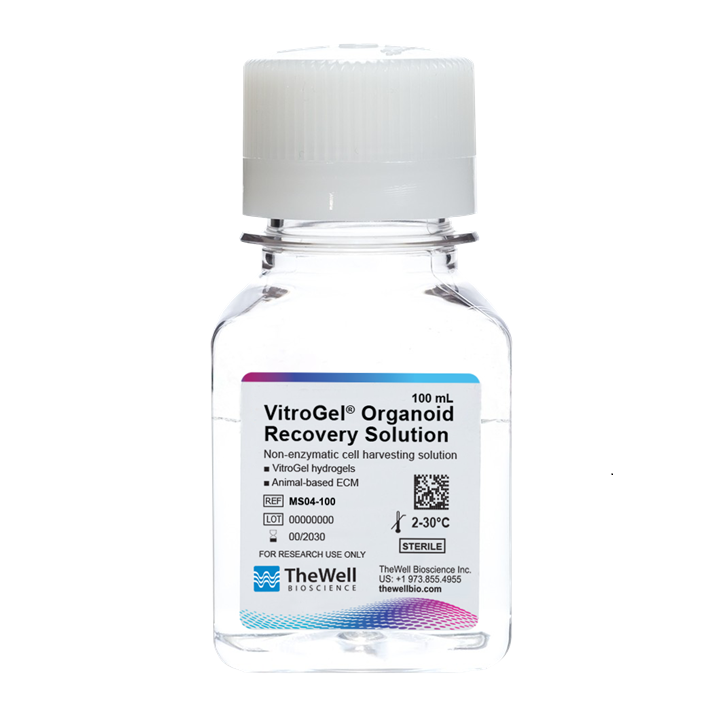
VitroGel® Organoid Recovery Solution is a non-enzymatic cell/organoid harvesting solution for quick and efficient recovery of 3D cells or organoids cultured with either VitroGel® hydrogels or an animal-based ECM.

The Cyto3D® Live-Dead Assay Kit is a quick one-step staining procedure for analysis on a dual-fluorescence system and versatile live/dead assay for 3D and 2D Cell Culture, Organoids, Spheroids, Stem Cells, and Fluorescence Microscopy.
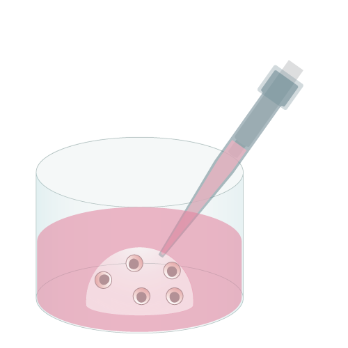
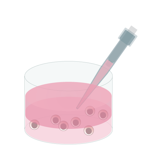
VitroGel® ORGANOID provides flexible options for culturing organoids, supporting a variety of methods to suit diverse research needs. It can be used for 3D cell encapsulation, 2D hydrogel coating, or creating hydrogel droplets for organoid growth. Additionally, VitroGel® can adapt to classical dome methods while also enabling whole-well culture setups, offering researchers unparalleled versatility and ease of use in organoid culture systems.
Widely used method for organoid generation.
Encapsulate cells within the hydrogel for excellent mechanical support and cell-matrix interactions.
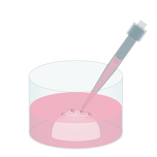
Add cells on top of hydrogel for optimal interactions among cells, hydrogel, and medium.
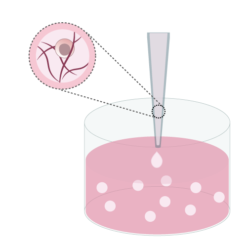
Create a Hydrogel-Cell droplet. This is a unique culture method only to the VitroGel® system for generating great cell-matrix interactions.
VitroGel® ORGANOID provides an optimal xeno-free environment for long-term culture, preserving the phenotypical properties of patient-derived organoids, tumoroids, and ex vivo tissue explants for weeks or even months. This enables reliable disease modeling, personalized medicine, and drug screening with high reproducibility.

A novel preclinical model of the
normal human breast
VitroGel® helps to maintain viable organotypic breast tissue cultures over 7 days without losing phenotype properties. Tissue maintains expression of estrogen and progesterone receptors and remains responsive to hormones.
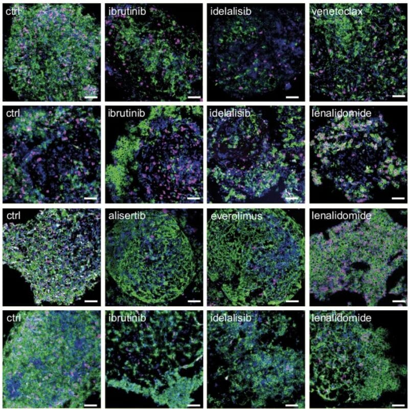
Development of patient-derived lymphomoids with preserved tumor architecture for lymphoma therapy screening
VitroGel® contributed significantly to the results by ensuring that the tissue fragments retained their histological and molecular features, allowing for effective testing of various clinically approved drugs.
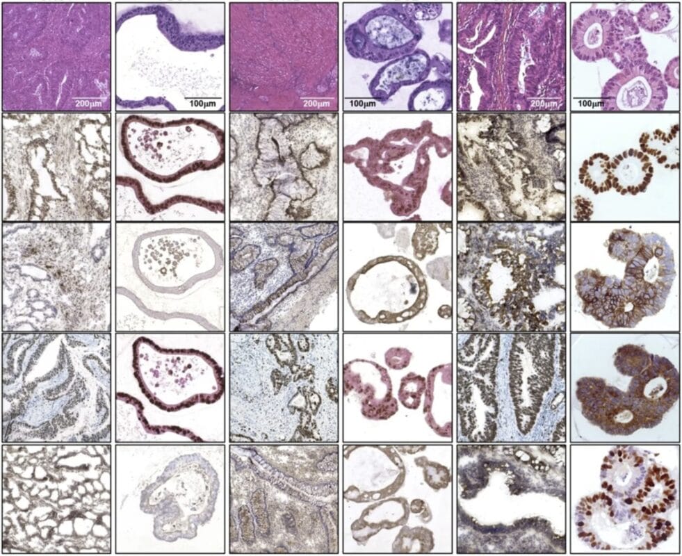
Harnessing Patient-Derived Organoids to Decode Immunotherapy Responses in Colorectal Cancer
VitroGel® revolutionizes colorectal cancer research by creating a xeno-free platform for patient-derived organoids, enabling groundbreaking insights into tumor-immune interactions and immunotherapy responses.
Designed to support both 2D maintenance and 3D scale-up, VitroGel® ORGANOID offers a complete xeno-free culture platform for iPSC-derived organoids. Providing a physiologically relevant matrix facilitates efficient differentiation, expansion, and tissue maturation, opening new possibilities of iPSC-derived organoids for tissue regeneration and regenerative medicine.
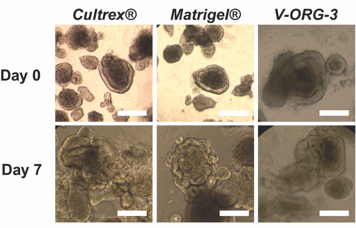
Intestinal Organoid Generation on animal-derived ECM vs. Xeno-Free VitroGel®
VitroGel® ORGANOID, Matrigel®, and Cultrex® equally facilitate the maturation of intestinal organoids. V-ORG-3 leads to larger and more mature hIO suggesting that the physical properties of xeno-free hydrogels can be harnessed to optimize organoid generation using these systems.
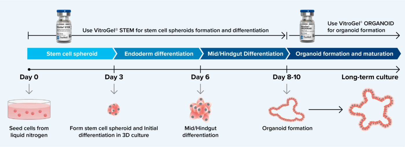
Xeno-free Organoid Generation Workflow for Stem Cell Spheroids Using VitroGel® STEM and VitroGel® ORGANOID Hydrogel System
VitroGel® ORGANOID is also able to effectively promote further organoid growth and development, allowing for long-term, high-quality cultures of these organoids. VitroGel® STEM and VitroGel® ORGANOID effectively streamline the organoid differentiation process, increasing the ease of generating organoids for a variety of applications.
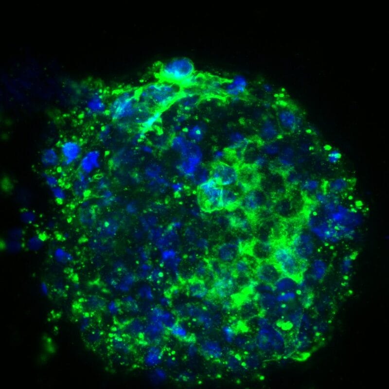
VitroGel® ORGANOID enables a seamless co-culture of organoids with immune cells, supporting immune cell penetration and interaction with organoids/tissue fragments within the matrix. Its optically clear matrix allows for high-resolution imaging and live-cell analysis, making it an ideal platform for studying tumor-immune interactions, immunotherapy applications, and host-microbiome interactions in a physiologically relevant 3D environment.
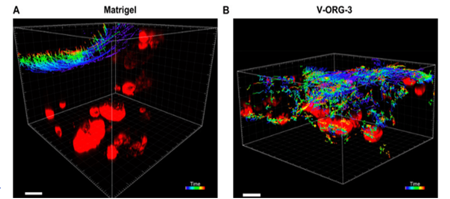
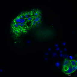
Establishing the First Co-Culture Model of Human Gastric Organoids and Dendritic Cells Using the Xeno-free VitroGel® ORGANOID Hydrogel System
DC migratory activity in Matrigel vs. VitroGel® ORG-3
Poor movement of MoDCs (green) when co-cultured with and HGOs (red) embedded in Matrigel. Improved migration of MoDCs (green) towards HGOs (red) embedded in V-ORG-3.
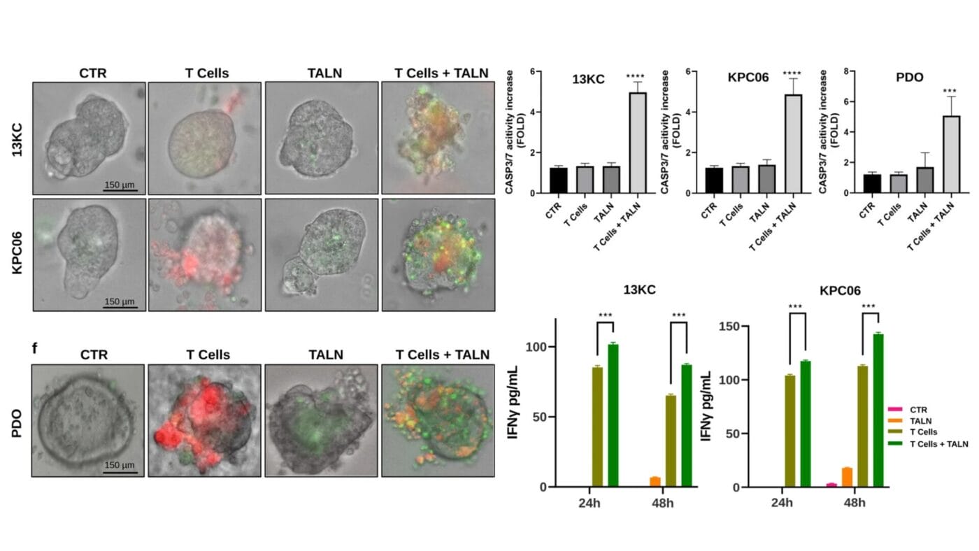
3D Organoids and Talniflumate: A Powerful Duo Against Pancreatic Cancer
In vitro recognition platform between Telomerase specific T cells and 3D-pancreatic cancer cultures from 13 KC and KPC06 mouse model. T cells are stained with CMPTX (red), and Caspase 3/7 activity is shown in green.
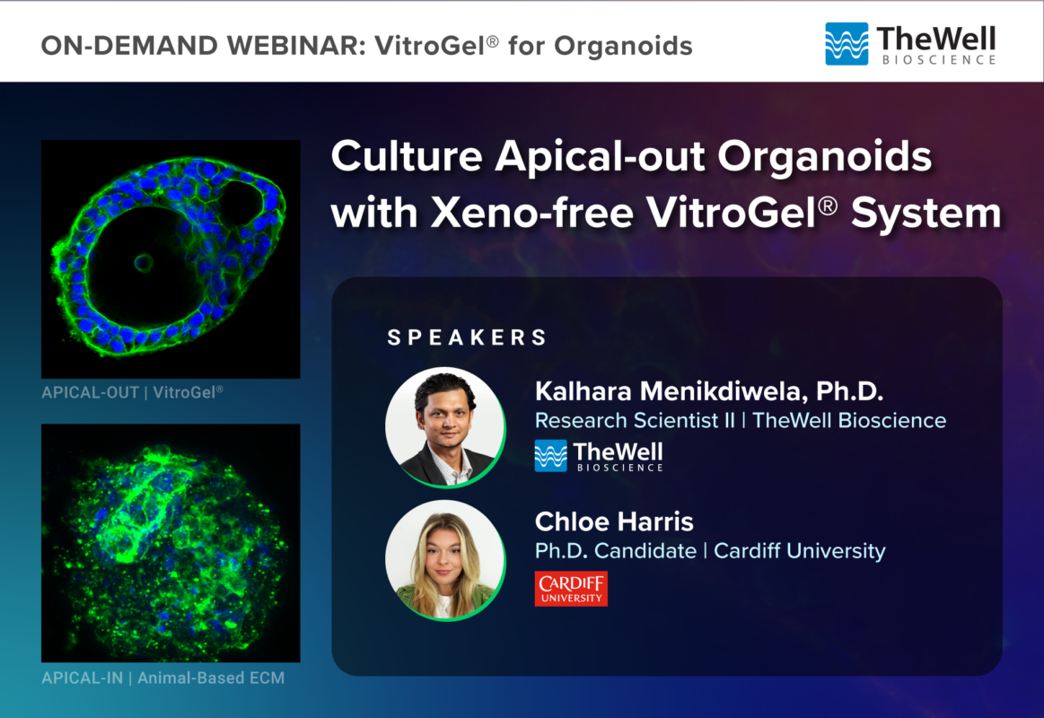
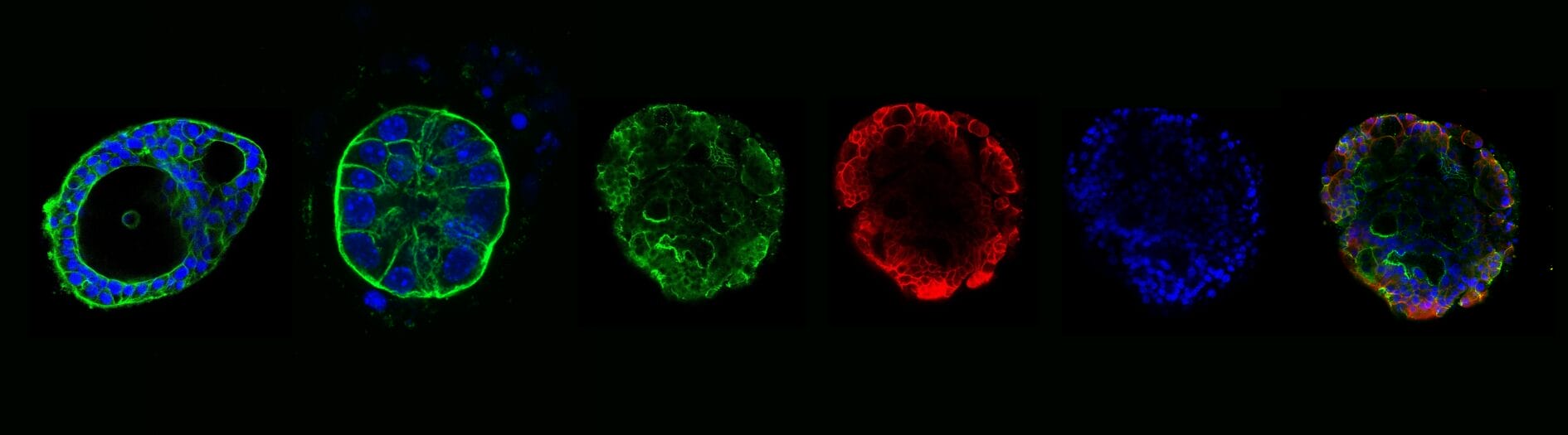
The polarity of organoids plays a vital role in overall development and functionality of organoids. Traditionally, organoids cultured in animal-based extracellular matrices (ECMs), such as Matrigel, often exhibited apical-in polarity. This limitation hinders their application in critical research areas, including host-drug interactions, intestinal host-microbiome studies, and nutrient uptake through epithelial cells, etc. However, organoids cultured in VitroGel® exhibit apical-out polarity, providing direct access to the apical side of the organoids to carry out advanced research which is not possible with apical-in organoids.
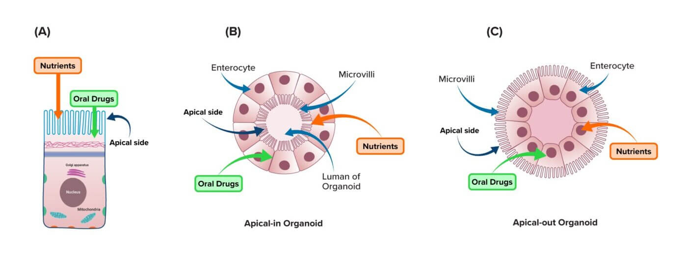
Fig. 1: Organoids culture in VitroGel® ORGANOID supports apical-out polarity. A. Nutrients and drugs are directly interacting with enterocytes at the apical-side. B. An organoid culture in animal-based hydrogel e.g. Matrigel with apical-in polarity. C. An organoid cultured in VitroGel® ORGANOID with apical-out polarity.
APICAL-OUT | VitroGel®
APICAL-IN | Matrigel®
VitroGel® ORGANOID supports apical-out polarity of organoid, in contrast to the apical-in polarity observed in animal-based hydrogels.
Organoids cultured in VitroGel® exhibit apical-out polarity, in contrast to the apical-in polarity observed in animal-based hydrogels. Furthermore, organoids cultured in VitroGel® demonstrated stable and controlled growth over time, unlike the fast, uncontrolled organoid growth (recapitulating growth patterns similar to tumors) commonly seen in animal-based hydrogels.

Long-term (60+ days) organoid culture with xeno-free VitroGel® ORGANOID. VitroGel® ORGANOID supports the development of intestinal organoids while maintaining structural and morphological integrity for over 60 days.
Mature long-term organoid culture support with xeno-free VitroGel® system:
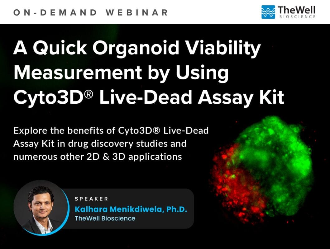
The Cyto3D® Live-Dead Assay Kit offers a quick and simple one-step staining procedure that accurately determines cell viability in both 3D and 2D cell cultures, making it highly versatile for various research applications. Its ability to provide even staining with clear resolution and no background interference enhances imaging quality and experimental precision, particularly for organoids, spheroids, and stem cells.

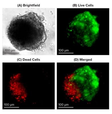
A Quick Organoid Viability Measurement by Using Cyto3D® Live-Dead Assay Kit
Live-dead cell viability images: Intestinal organoids stained with Cyto3D® Live-Dead Assay Kit. A bright field image of a mature intestinal organoid. Images show live cells (B: Green) and dead cells (C: Red) in a mature intestinal organoid.
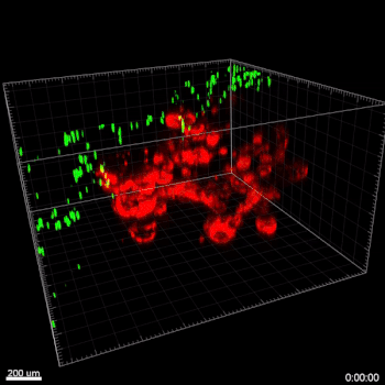
Use of Cyto3D® Live-Dead Assay Kit to test cell viability in 3D cultured Patient Derived Organoids (PDOs)/ovarian tumors.
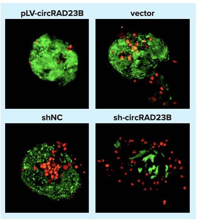
Functions of circRAD23B in patient-derived organoids (PDOs). The effects of 40 µM carboplatin pressure on PDOs for 48 hours and organoids stained with Cyto3D® reagent indicate cell viability/death; live cells are green, while dead cells are red.
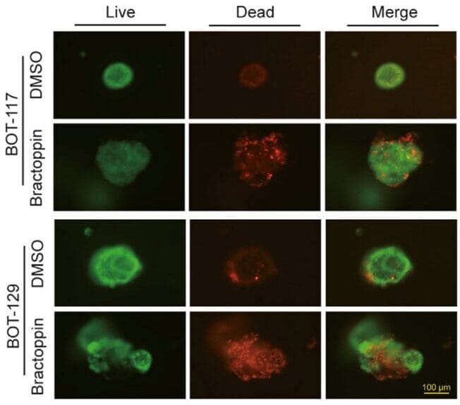
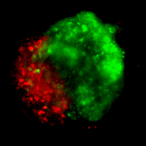
Advancing Borderline Ovarian Tumor Research: Precision Viability Assessment in Patient-Derived Organoid Model
Cyto3D® Live-Dead Assay Kit enables the assessment of viability in patient-derived borderline ovarian tumor organoids for the evaluation of novel therapeutic compounds.
Adding hydrogel as a dome may cause the gel floating issue. Therefore, we don’t recommend the dome method; however, some customers still want to adopt the dome method. At this point, the non-tissue culture-treated plate can hold the dome better than the TC-Plate with some tips. You may need to add the dome as soon as you mix the gel and cells. Also, warming up the medium can help a little (slow down the gel formation’s speed).
However, the dome method is not 100% successful with non-TC plates, and some scientists really have a hard time with it. That is why we recommend covering the whole bottom of the well with our hydrogel. We usually use a 48 or 96-well plate (about 100-150 uL per well of a 48-well plate or 50 uL per 96-well plate). For full bottom coverage, the TC plate is better. Alternatively, you could coat the plate with 0.1-0.5% gelatin for 10 minutes and then remove the gelatin before adding the hydrogel.
For the dome method,
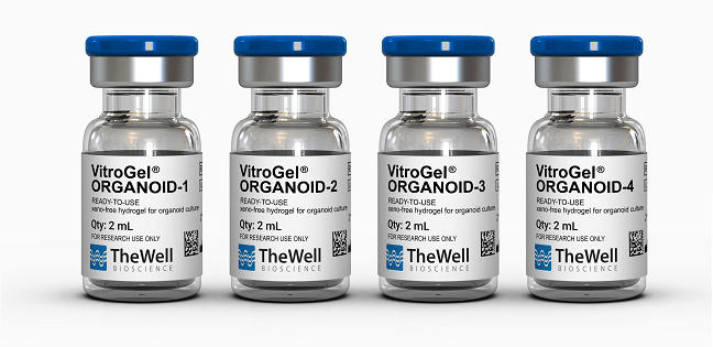
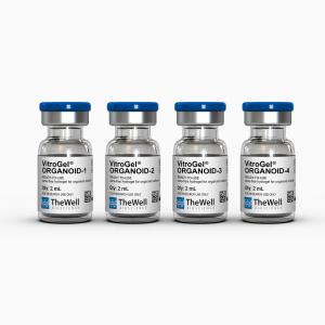
Because there is a wide range of organoid cell resources from stem cells, patient-derived tissue, co-culture, and PDX, it is hard to tell which organoid hydrogel would perform the best for the researcher’s experiment. Therefore, the VitroGel® ORGANOID Discovery Kit helps researchers screen the four different formulations to determine the best organoid hydrogel version for your organoid conditions.
The VitroGel® ORGANOID Discovery Kit includes the four different formulation types of VitroGel® ORGANOID hydrogels in 2mL :
Each of the four types has various bio-functional ligands, mechanical strengths, and degradability to fulfill the needs of different organoid culture conditions. From our findings, versions 1, 2, or 3 are suitable for gastric organoids. Versions 1 and 3 are suitable for lung organoids. Version 2 or 3 are suitable for brain organoids, and versions 3 or 4 are suitable for cancer organoids. By saying that, we still suggest using the Discovery Kit to screen the optimal hydrogel for your organoid conditions quickly.
The VitroGel® ORGANOID has four different versions, each formulated with various biofunctional ligands, mechanical strengths, and degradability to fulfill the needs of different organoid culture conditions.
The VitroGel® ORGANOID-3 would be a good starting point as many researchers get good results on this hydrogel for their organoid culture.
Hydrogels - Ready-To-Use
ready-to-use, xeno-free (animal origin-free) hydrogel system for organoid culture
Hydrogels - Ready-To-Use
ready-to-use, xeno-free (animal origin-free) hydrogel system for hPSCs 3D static suspension culture and scale-up

Cell Harvesting Solution
Non-enzymatic cell harvesting solution to recover cells/organoids from hydrogel or an animal-based ECM within 15 minutes. Improved formulation over VitroGel® Cell Recovery Solution.

Biomarkers
Versatile, live/dead cell viability analysis for 3D and 2D cell culture. IN STOCK