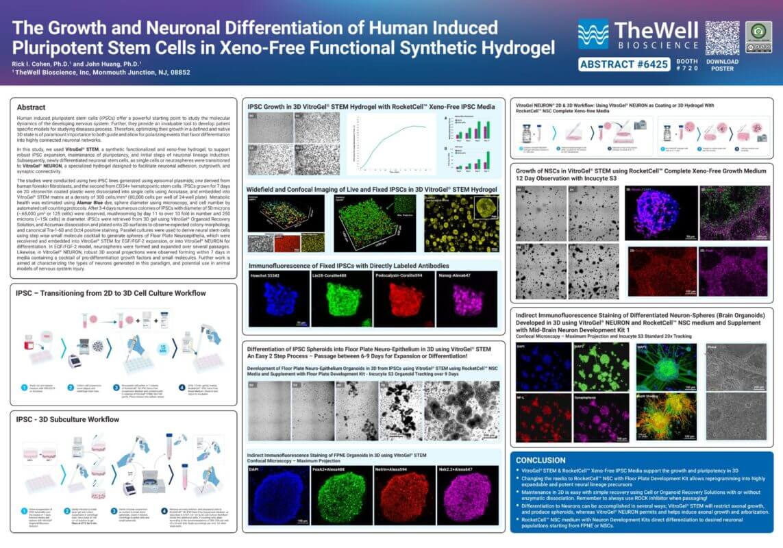
NEW
VitroGel® NEURON
Ready-to-use, xeno-free hydrogel system for neural stem cell and neuron cultures.
VitroGel® NEURON for 3D Neuron Culture
Xeno-free and biofunctional
100% synthetic. Animal & human origin-free hydrogel. No growth factors or proteins in the gel solution.
Room temperature operation
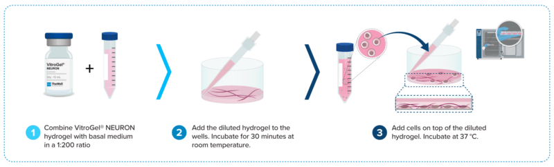
Stable at room temperature. Easy handling. Gelation is not temperature dependent. Learn how gelation works >

Supports 3D and 2D neuronal cell culture
Suitable for 3D and 2D culture of immortalized neuronal neuroblasts, iPSC-derived NSCs, neuron, and culturing of neural stem cells (NSCs) as single cells or neurospheres.

Injectable hydrogel for cell therapy
Its unique injectable properties make it an effective carrier for neuron-based cell therapy, spinal cord injury repair, and other regenerative medicine applications.
VitroGel® NEURON is the first-of-its-kind synthetic, xeno-free hydrogel specifically engineered to support both 2D and 3D neuronal culture and in vivo delivery. This groundbreaking platform eliminates the variability of animal-derived ECMs, such as Matrigel®, and provides a defined, reproducible, and scalable environment for neuronal growth, differentiation, and network formation.
With its ready-to-use, room-temperature-operation formulation, VitroGel® NEURON streamlines workflows, is transparent, and imaging-friendly, enabling real-time visualization of neural development. The system is optimized for primary cells, cell lines, iPSC-derived neural stem cells, and neuron maturation in both 2D and 3D formats, making it a powerful tool for translational neuroscience research.
By combining a precision bio-functional microenvironment and injectable-friendly features, VitroGel® NEURON bridges the gap between basic research and future biomedical applications.
Superior for 2D Thin Coating over Animal-based ECM

200X Dilution
4X more usage than animal-based ECM.

Long-Term Culture
Over 4 weeks of maintenance and
post-differentiation culture.

Easy and Fast Operation
No need to remove the coating solution,
just seed cells directly.
3D Neuron Culture Made Easy with VitroGel® NEURON

3D Neuron Differentiation
Supports both single cells and neuron spheroids.

Excellent Cell-Cell Communication
Enhanced cell extension and cell-matrix interactions.
Imaging-Friendly
Transparent and direct staining within the hydrogel for high-quality image data.
Enhance your NSC workflow with these products
- RocketCell™ 3D NSC Xeno-Free Complete Growth Kit↗
An all-in-one, ready-to-use kit for 3D neural stem cell culture. - RocketCell™ NSC Xeno-Free Complete Growth Kit↗
An all-in-one, ready-to-use kit for 2D neural stem cell culture. - RocketCell™ NSC Xeno-Free Growth Medium↗
Cell culture medium for NSC culture
Specifications
| Formulation | Xeno-free, biofunctional hydrogel |
| Use | 3D and 2D neuron and neural stem cell culture |
| Operation | Ready-to-use at room temperature |
| Injection | Injectable hydrogel for in vivo studies and lab automation |
| pH | Neutral |
| Storage | Store at 2-8°C. Ships at ambient temperature |
| Sizes | 10 mL and 2 mL |
Recommended Products
VitroPrime™ Spread-Attach Plate, combined with VitroGel® NEURON, supports exceptional 2D and 3D neuron growth. No PDL-coated plates required.
VitroPrime™ Spread-Attach Plate

Data and References
2D THIN COATING METHOD
2D: CASE 1
2D NSC Maintenance (1:200 coating on VitroPrime™ Spread-Attach Plate)
A
B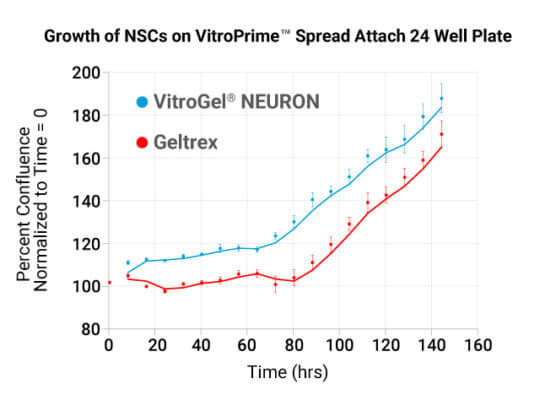
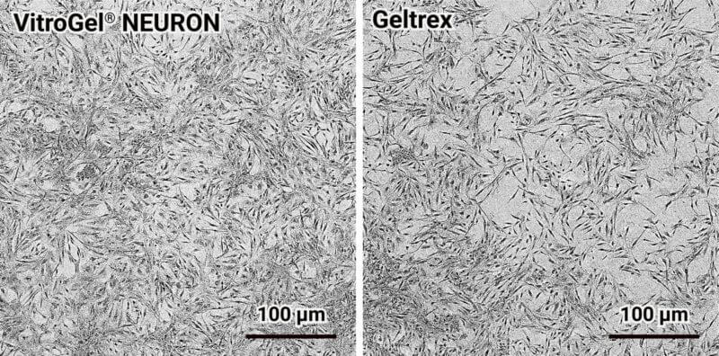
Figure 1. VitroGel® NEURON 2D Thin Coating for Growth of iPSC-Derived NSCs and Confluence Analysis on VitroPrime™ Spread-Attach Plates.
A. iPSC-derived NSCs (CD34-eIPSC-NSC) were plated at 50,000 cells per well on 24-well VitroPrime™ Spread-Attach Plates coated with either VitroGel® NEURON (1:200) or Geltrex®, and cultured for 6 days. Images were captured using the Incucyte® S3 system. B. NSC growth was quantified by percent confluence over time, with images collected every 8 hours and analyzed relative to the initial timepoint (T = 0 + 4 hrs).
2D: CASE 2
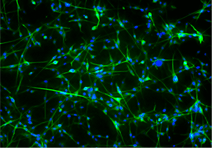
2D B35 Neuronal Neuroblast Differentiation (2D thin coating method)

Figure 2. 2D thin coating method using VitroGel® NEURON hydrogel promotes B35 neuronal neuroblast differentiation. Immunofluorescence staining of neuronal cultures was performed to evaluate the presence of the neuron-associated marker beta-III-tubulin on day 7 post-differentiation induction. The cultures were visualized with the Keyence BZ-X system.
2D: CASE 3
2D NSC Dopaminergic Differentiation
A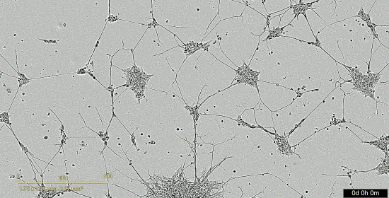
Figure 3. 2D thin coating with VitroGel® NEURON hydrogel supports the development of Dopaminergic Neuron Progenitors (DNPs)
A. Video shows robust survival of DNP colonies, and elaboration of neurites; B & C. IncuCyte Neurotrack analysis software was used to quantify the expansion of the area of DNP colonies, and their enhanced complexity of neurite elongation.
B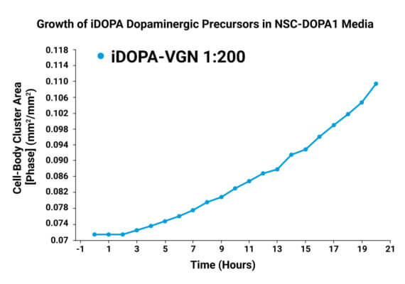
C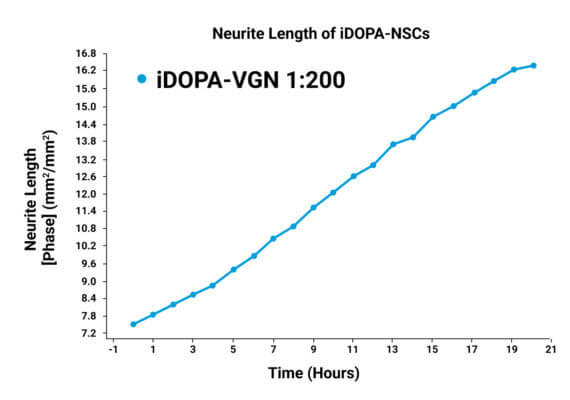
2D HYDROGEL COVERING “BLANKET METHOD”
![]()
2D BLANKET: CASE 4
2D B35 Neuronal Neuroblast Differentiation (2D “blanket” method)
![]()
Figure 4: 2D “Blanket” method using VitroGel® NEURON hydrogel sustains in vitro neuronal differentiation. Immunofluorescence staining of neuronal cultures was performed to evaluate the presence of the neuron-associated marker beta-III-tubulin on days 7, 14, 21, and 30 post-differentiation induction. A. Light microscopy image showing neuronal cultures. B. Green-fluorescent image illustrating the presence of beta-III-tubulin. C. The nuclei were observed using DAPI (blue) staining. D. Combination of B and C images. The cultures were visualized using the Molecular Devices Image Xpress Nano system at a 20X magnification.
2D BLANKET: CASE 5
NSC Spheroids Differentiation (2D “blanket” method)

3D CELL CULTURE
3D: CASE 1
Long-term 3D neuronal differentiation (B35 cells)
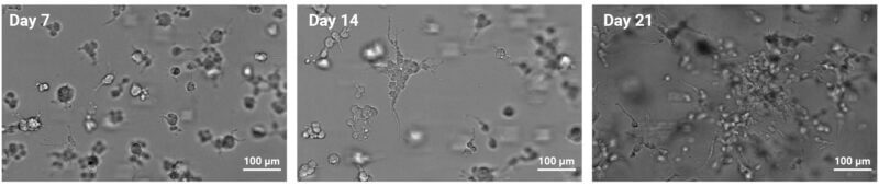
Figure 5. VitroGel® NEURON sustains long-term 3D neuronal differentiation.
Bright field microscopy images depicting 3D B35 neuronal neuroblast differentiation in VItroGel® NEURON on days 7, 14, and 21. The images were obtained with the Keyence BZ-X microscope.
3D: CASE 2
3D NSC culture with VitroGel® NEURON as single cell encapsulation
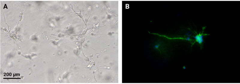
Figure 6. 3D encapsulation culture of NSC cells as a single cell suspension in VitroGel® NEURON.
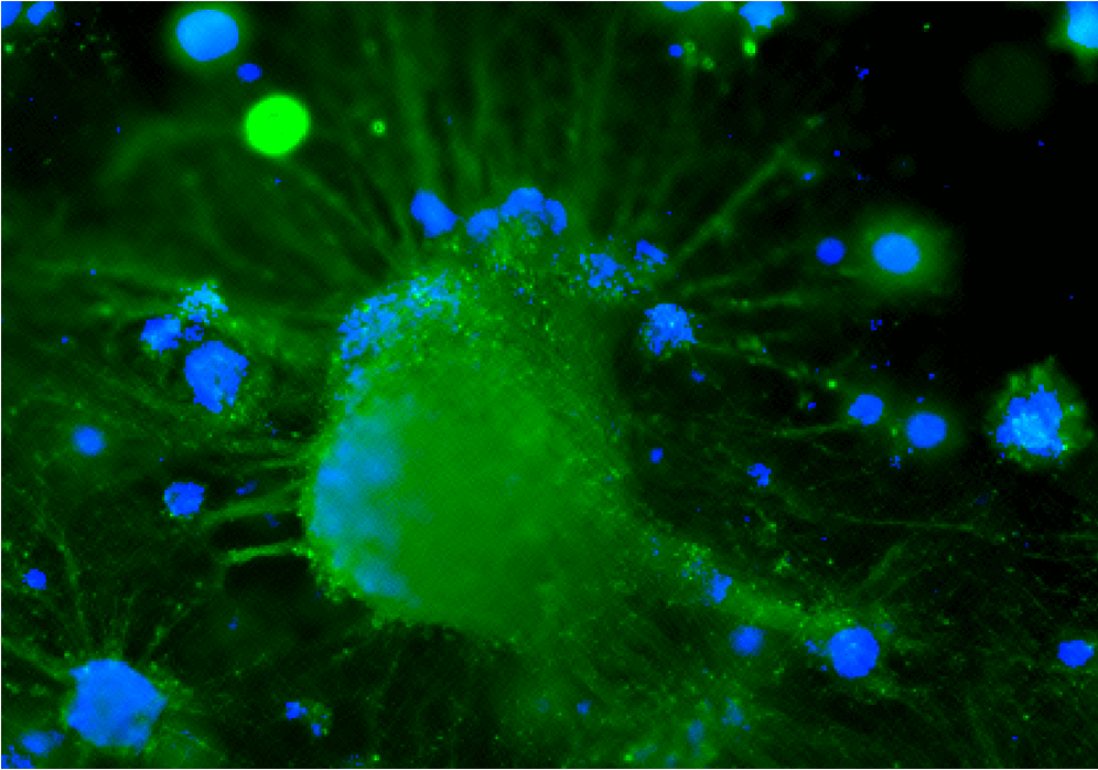
3D NSC cultures were established by preparing a single-cell NSC suspension with RocketCell™ NSC Xeno-Free Medium and mixing with VitroGel® NEURON for 6 days. A. Light microscopy images of NSC cultures demonstrating that VitroGel® NEURON Hydrogel and RocketCell™ NSC Xeno-Free Media Support neurite outgrowth. B. Immunofluorescence staining was performed to evaluate the presence of beta-III-tubulin, a neuron maturation marker. The image was obtained with the Keyence BZ-X microscope at a 20X magnification.
3D: CASE 3
3D Development of Dopaminergic Neurons from NSC spheroids
A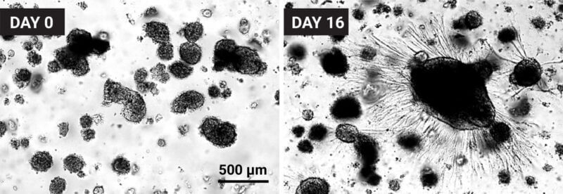
B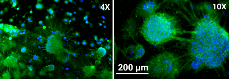
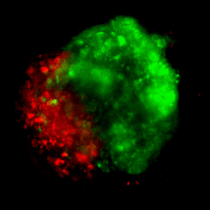
Figure 7. VitroGel® NEURON hydrogel supports the 3D development of dopaminergic neurons.
iPSCs were 3D cultured in VitroGel® STEM with RocketCell™ IPSC Xeno-Free Growth Medium (Cat. No. RC02-GM) for 6 passages and were differentiated into Floor Plate Neuro-Epithelial Spheroids. The spheroids were recovered from the hydrogel using the VitroGel® Organoid Recovery Solution and embedded in VitroGel® NEURON.
A. Brightfield images of spheroid on day 0 (A, left) that were differentiated for 16 days (A, right). B. The samples were fixed and stained with beta-III-tubulin antibody and DAPI, depicting that the majority of the spheroids are neurons with the ability to form axonal connections. The cultures were visualized with the Keyence BZ-X microscope at 4X and 10X magnifications.
3D: CASE 4
3D Encapsulation
Culture of NSCs
Figure 8. 3D encapsulation culture of NSCs in VitroGel® NEURON hydrogel as NSC spheroids with zonal projections.
iPSCs were cultured and expanded using the RocketCell™ 3D iPSC Xeno-free Complete Growth kit. The cells were harvested from the hydrogel with the VitroGel® Organoid Recovery Solution and embedded in VitroGel® NEURON to induce iPSC differentiation into NSCs with CytoGrow™ Growth factors. The NSCs were differentiated into dopaminergic neurons with the RocketCell™ NSC Neuron Differentiation Kit.
3D: CASE 5
3D Differentiation of Cholinergic Neurons
Figure 9. Development of Cholinergic Neurons from IPSCs using FPNE Protocol using VitroGel® NEURON
FPNE Organoids were recovered from VitroGel® STEM using Organoid Recovery Solution and plated with VitroGel® NEURON in RocketCell™ NSC Media and Supplement with RocketCell™ Neuron Maturation Kit cytokines. Cultures were developed for 14 days prior to indirect immunostaining for Phosphorylated Neurofilament (RT97, purple) and Choline Acetyl Transferase (CHAT, green).
Enhance your neuron cell culture with these products:
New
Cell Culture Medium (Xeno-Free)
All-in-one, ready-to-use kit for 3D neural stem cell culture.
New
Cell Culture Medium (Xeno-Free)
All-in-one, ready-to-use kit for 3D and 2D neural stem cell culture.
New
Cell Culture Medium (Xeno-Free)
Xeno-Free complete medium for 3D and 2D neural stem cell culture
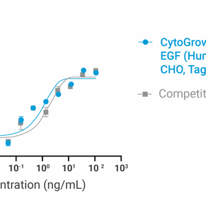
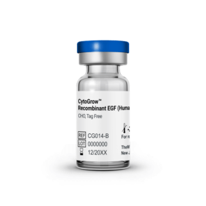
CytoGrow™ - Recombinant Proteins
Premium CytoGrow™ growth factor suitable for cell culture supplementation, wound healing research, epithelial biology, and cancer signaling studies.

3D Culture Vessels
Unique surface treated for superior hydrogel spreading, adherence, and uniform surface.
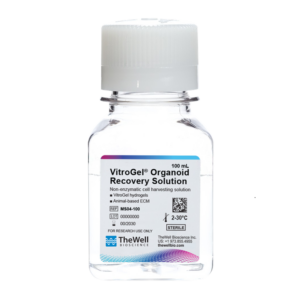
Cell Harvesting Solution
Non-enzymatic cell harvesting solution to recover cells/organoids from hydrogel or an animal-based ECM within 15 minutes.

Downstream Cell Analysis
Fast (15 min), versatile, live/dead cell viability analysis for 3D and 2D cell culture. IN STOCK
| Size | 2 mL, 10 mL |
|---|
Related products
Hydrogels - Ready-To-Use
ready-to-use, xeno-free (animal origin-free) biofunctional hydrogel system

Hydrogels - Ready-To-Use
ready-to-use, xeno-free (animal origin-free) hydrogel system for organoid culture
Hydrogels - Ready-To-Use
ready-to-use, xeno-free (animal origin-free) hydrogel system for hPSCs 3D static suspension culture and scale-up
Hydrogels - Ready-To-Use
ready-to-use, xeno-free (animal origin-free) hydrogel system for 3D culture of HEK293 cells
NEW

3D Xeno-Free Hydrogels
Xeno-free hydrogel that supports spheroid formation, tumoroid culture, and uniquely enables EMT biology Full xeno-free EMT kit available. Click here
Hydrogels - Ready-To-Use
ready-to-use, xeno-free (animal origin-free) hydrogel system for mesenchymal stem cell 3D culture and scale-up