
High cell quality
Yields high-quality 3D stem cells with high success rate for downstream differentiation.
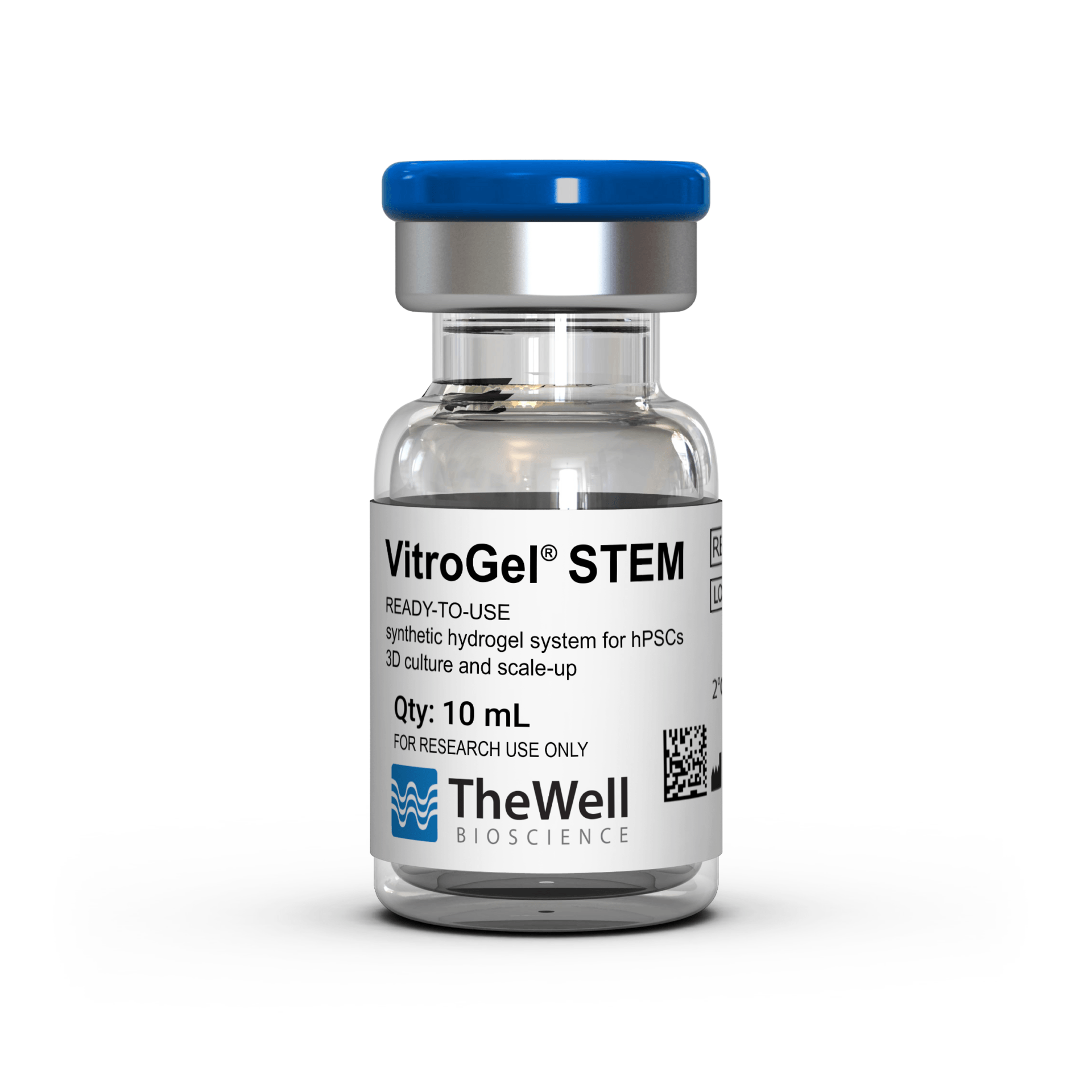
VitroGel® STEM
Ready-to-use, synthetic, chemically defined hydrogel system for hPSCs 3D static suspension culture and scale-up
VitroGel® STEM
Ready-to-use, synthetic, chemically defined hydrogel to support both scale-up and 3D differentiation of hPSCs

Synthetic, Chemically Defined Hydrogel
100% synthetic. Animal & human origin-free, biofunctional and biocompatible hydrogel.

High Expansion rate
Faster growth rate than traditional 2D culture and yields uniform spheroid sizes.

5-min. protocol
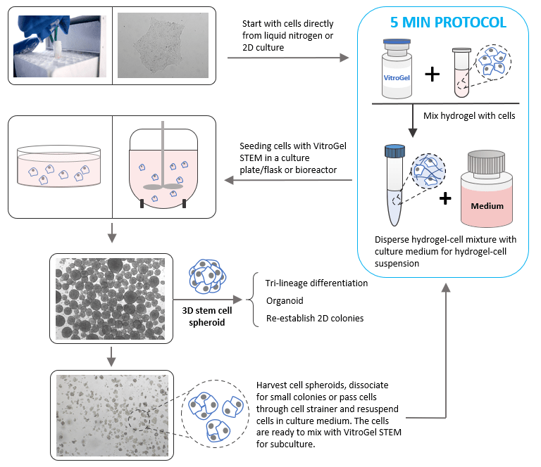
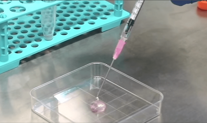
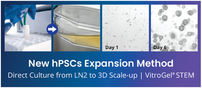
Establishes 3D stem cell expansion directly from liquid nitrogen with simple mixing steps, no matrix coating required.

Easy cell harvesting
Simple and efficient cell harvesting by either centrifuge or the non-enzymatic VitroGel® Organoid Recovery Solution.

Flexible and compatible
Compatible with most hPSC culture media and tissue culture vessels.
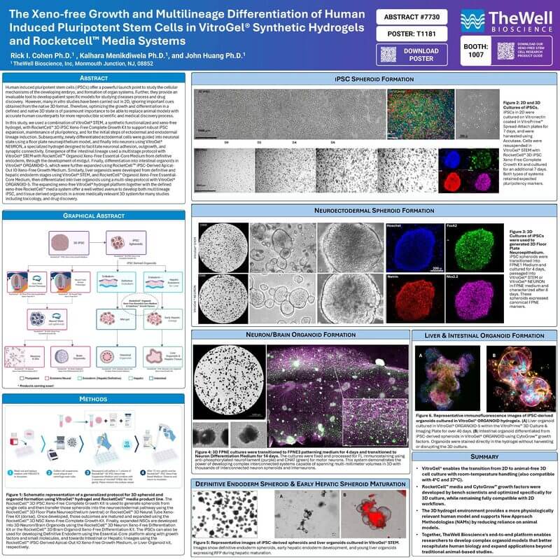
VitroGel® STEM is a synthetic, chemically defined hydrogel system developed to support scale-up and 3D differentiation of human pluripotent stem cells (hPSCs) to create a high-throughput system to model various tissue and disease states.
This hydrogel system is ready to use with an optimized formulation that fully supports the rapid expansion of high-quality 3D stem cell spheroids with pluripotent properties. hPSCs directly thawed from liquid nitrogen or passaged from 2D matrix-coated culture vessels can be immediately mixed with the hydrogel solution for static suspension cultures. Moreover, the optimization protocol is ideal for time-sensitive experiments, as it does not require excessive medium exchanges, which can ultimately save time and materials. This hydrogel system is compatible with most hPSC culture media and tissue culture vessels. The 3D stem cell spheroids developed using this system can be used for further sub-cultures, patterned differentiation, organoid development, or re-establishing 2D culture morphologies.
Enhance your stem cell culture research with the following animal origin-free growth kits:
- RocketCell™ 3D iPSC Complete Growth Kit, AOF↗
An all-in-one optimized kit for 3D expansion of pluripotent stem cells - RocketCell™ iPSC Complete Growth Medium, AOF↗
Animal origin-free medium to support the expansion of pluripotent stem cells
“Just add cells” 5 min protocol. No matrix coating required.
VitroGel® STEM is ready-to-use. Just mix it with your hPSCs. No laborious matrix coating is required to maintain and expand your stem cells.
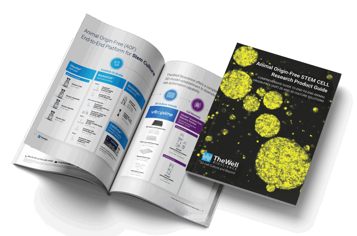
Complete Guide for
Animal Origin-Free Stem Cell Culture
This comprehensive product guide features validated applications, optimized workflows, and integrated solutions designed to support reproducible cell expansion, maintenance, and differentiation across a wide range of stem cell research applications.
Specifications
| Formulation | Synthetic, chemically defined hydrogel |
| Use | 3D static suspension culture for hPSCs |
| Operation | Ready-to-use at room temperature |
| Biocompatibility | Biocompatible, safe for animal studies |
| Injection | Injectable hydrogel for in vivo studies and lab automation |
| Cell Harvesting | VitroGel Organoid Recovery Solution 5-15 min cell recovery |
| pH | Neutral |
| Storage | Store at 2-8°C. Ships at ambient temperature |
| Sizes | 10 mL and 2 mL |
| Number of Uses | (10 mL) 90-180 mL suspension culture (2 mL) 15-30 mL suspension culture |
Recommended Product
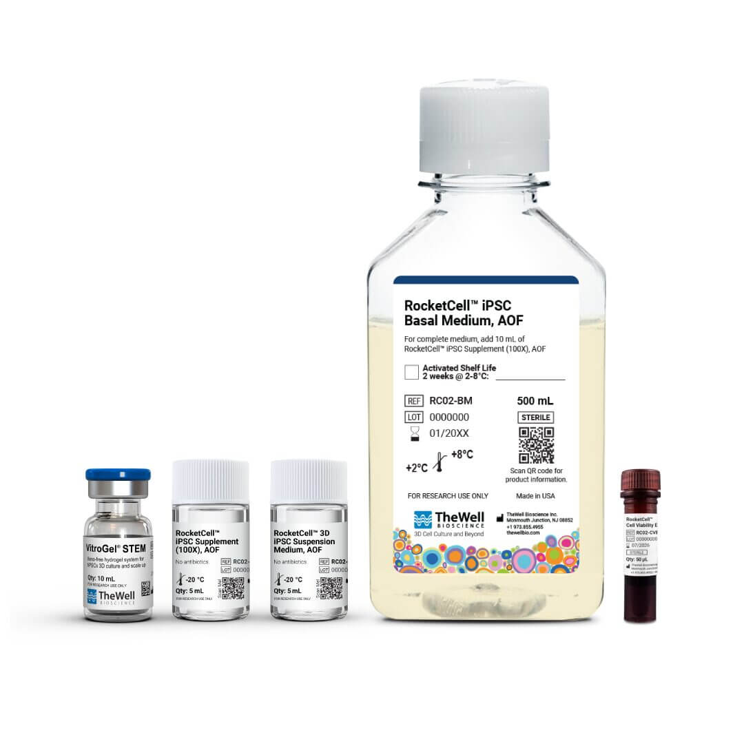
RocketCell™ 3D iPSC
Complete Growth Kit, AOF
An all-in-one optimized kit for 3D expansion of pluripotent stem cells.
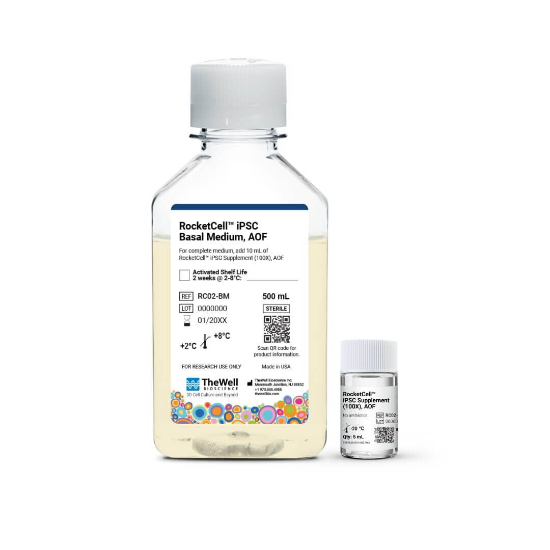
RocketCell™ iPSC Growth Medium, AOF
Animal origin-free medium for 2D and 3D expansion of pluripotent stem cells.


Comparison VitroGel® STEM vs Natural ECM
VitroGel® STEM | Natural ECM |
|
Culture Method | 3D Suspension | 2D Matrix Coating |
Synthetic, chemically defined | Yes | No |
3D Spheroid Formation | Yes | No |
3D Differentiation (directly) | Yes | No |
Easy to scale-up | Yes | No |
Preparation Time | < 10 mins | 2 hours + |
Cell viability after seeding | ●●●●● | ●●○○○ |
Cell Proliferation | ●●●●● | ●●●○○ |
Easy Cell Harvesting | ●●●●● | ●●○○○ |
Easy Cell Quality for Differentiation | ●●●●● | ●●○○○ |
Easy for Sub-culture | ●●●●● | ●●●○○ |
Data and References
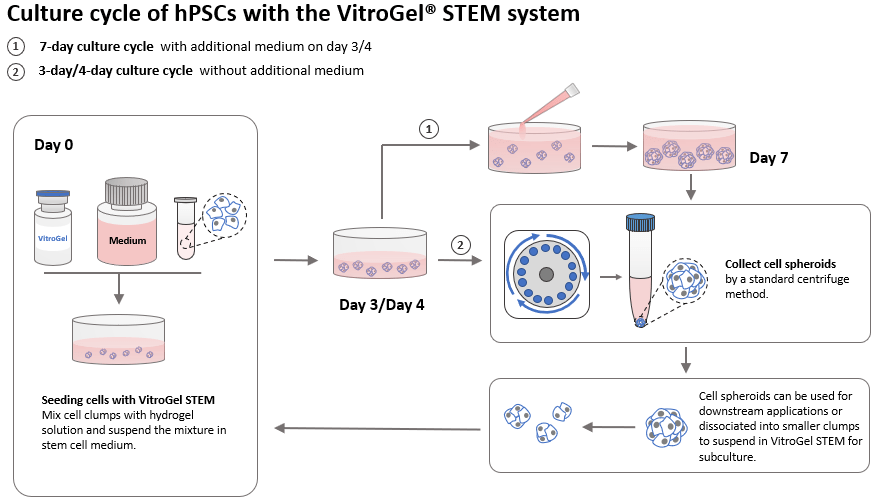
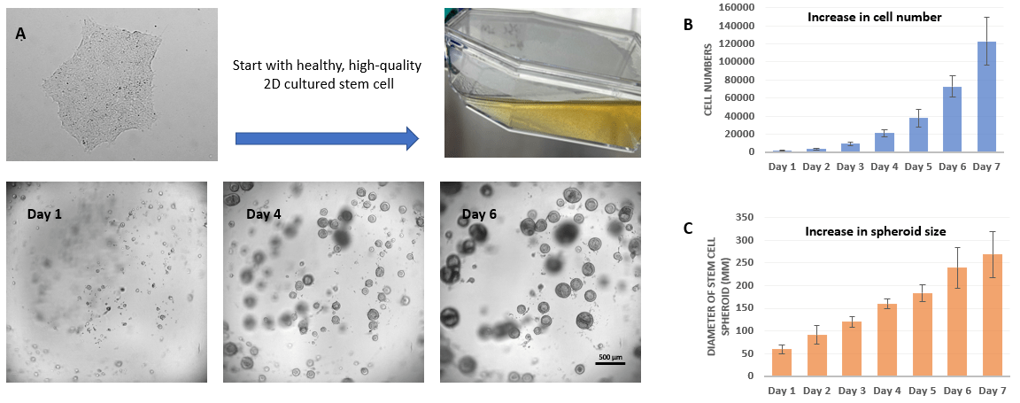 Figure 1. 3D static suspension culture of hPSC from 2D matrix culture
After 24 hours, small hPSC spheroids start to form. From day 1 to 6, cells in the suspension cultures quickly grow, leading to the generation of healthy and high-quality stem cell spheroids. After day 3, cell numbers grew exponentially (Figure 1B), and spheroid size steadily increases (Figure 1C). The hPSC spheroids display characteristics of shallow craters or pockmarks, indicating expression of hPSC markers and successful expansion of healthy and high-quality stem cell spheroids. The resulting spheroids provide researchers with large numbers of healthy hPSCs for further experiments.

Figure 2. 3D static suspension culture of hPSC directly from Liquid Nitrogen (LN2)
Start the suspension culture by using healthy and high-quality cells directly from LN2. hPSC-hydrogel aggregates successfully to form healthy spheroids after 1 day in culture. The hPSC spheroids continue to expand from day 1 to 6 (Figure 2A). The resulting hPSC spheroids also show hallmark features of healthy and high-quality stem cell spheroids, i.e., shallow craters or pockmarks. Figure 2B shows that hPSC static suspension cultures from liquid nitrogen are positive for Alkaline Phosphatase, indicating successful expansion of healthy stem cell populations.
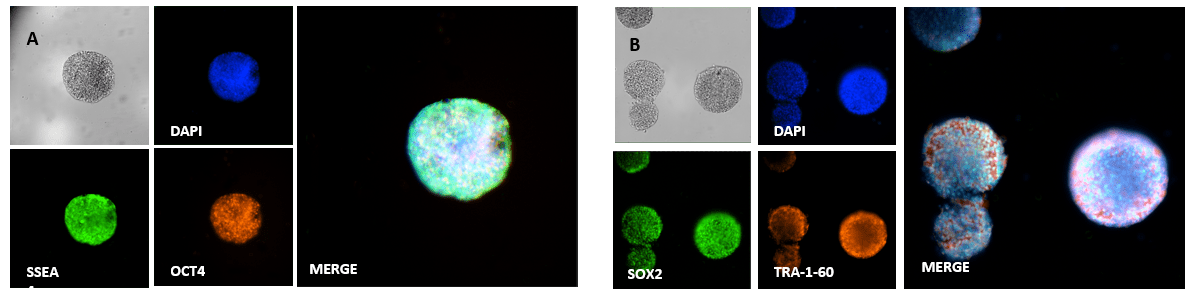
Figure 3. Immunofluorescence images of hPSC spheroids with key pluripotent stem cell markers
VitroGel STEM ensures the undifferentiated state of stem cell lines during scale-up. As shown in Figure 3, hPSC aggregates in VitroGel STEM hydrogel and retain pluripotency after 7 days, evidenced by the expression of key pluripotent stem cell markers, SSEA4, OCT4, SOX2, and TRA-1-60.
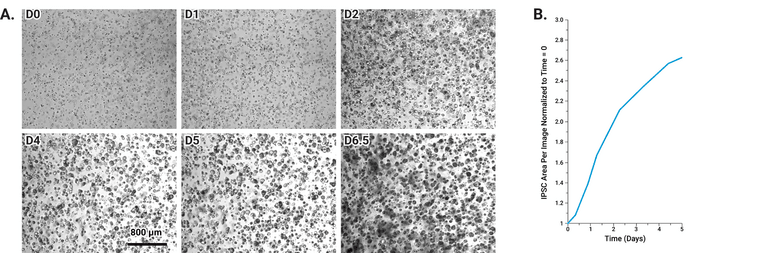
Figure 4. 3D Expansion of Human iPSCs
A: Representative time-lapse micrographs show human iPSCs transitioning from single cells and small aggregates into uniform 3D spheroids within the VitroGel® STEM hydrogel (seeded cells from a 2D source on Day 0). The spheroids exhibit consistent morphology and reach diameters of approximately 85–100 μm after several days in culture. B: Quantitative analysis of spheroid growth demonstrates a steady increase in total spheroid area over time, reaching an average of nearly 300% expansion after 6.5 days, confirming robust and reproducible 3D proliferation under animal origin-free culture conditions.
A.
B.
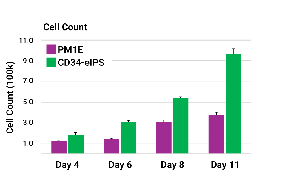
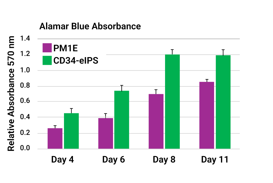
Figure 5. Quantitative Assessment of 3D iPSC Growth in VitroGel® STEM
A: Alamar Blue absorbance assay showing a progressive increase in metabolic activity of iPSCs cultured in 3D, indicating robust cell proliferation and viability over the culture period.
B: Corresponding cell count analysis confirming steady growth and expansion of iPSC spheroids within the VitroGel® STEM hydrogel, demonstrating the kit’s capability to support sustained 3D cell proliferation.
These findings demonstrate that metabolic assays, such as Alamar Blue, can be used with IPSCs embedded in VitroGel® STEM hydrogel to estimate cell expansion, compared with recovering cells from the hydrogel and using traditional cell counting methods. These figures highlight that IPSCs can respond differently to the 3D environment, so grow better than others. The overall pattern of the two assays are similar. and it is well known that Alamar Blue assays can saturate. We see that at Day 11 in the CD34-eIPS line more than other.
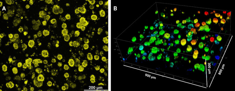
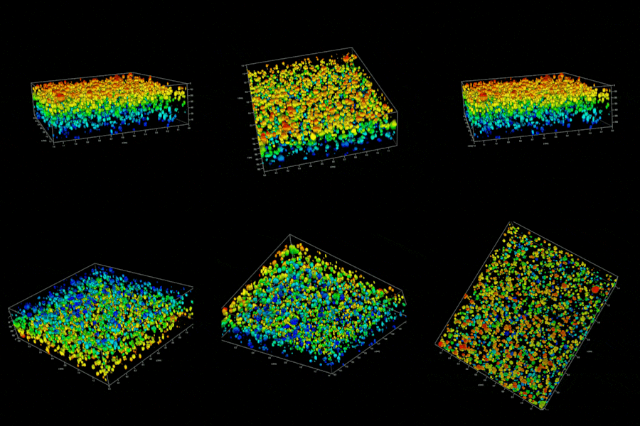
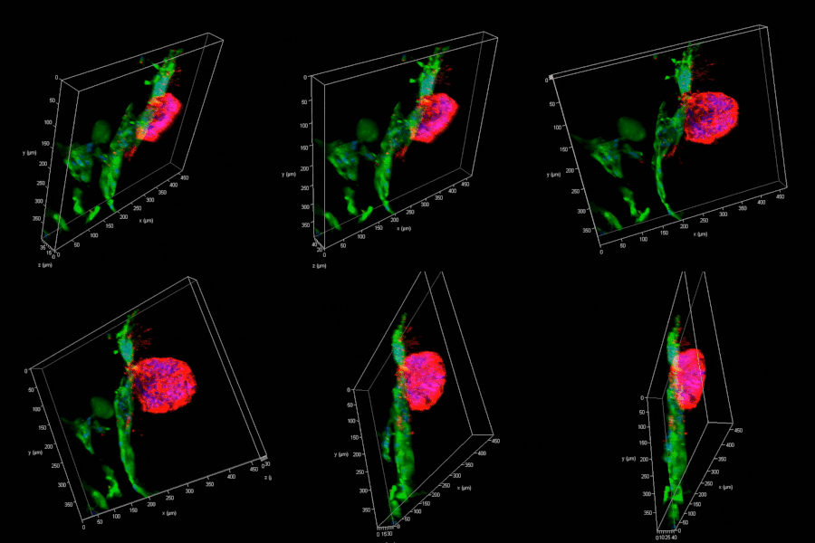
Figure 6: Live Cell Surface Immunofluorescent Staining of Human iPSCs Grown in VitroGel® STEM Hydrogel
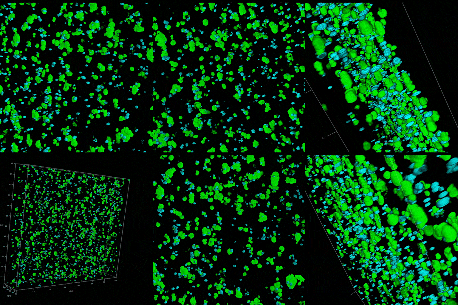
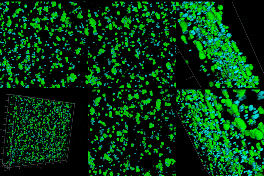
IPSCs (100k/well) from 2D sources were plated in 24-well VitroPrime™ 3D Culture and Imaging Plate. After 7 days, the medium was removed and replaced with media containing a diluted anti-EPCAM-PE-Alexa594-labeled antibody. The mixture was incubated for 1 hour at 37 °C, and then the well was rinsed three times with 5 min incubation with 0.5 mL of Growth Media. The well was imaged on a Leica MICA confocal microscope (A). The rendered image (B) is 300 microns deep, with a field approximately 800 x 900 microns.
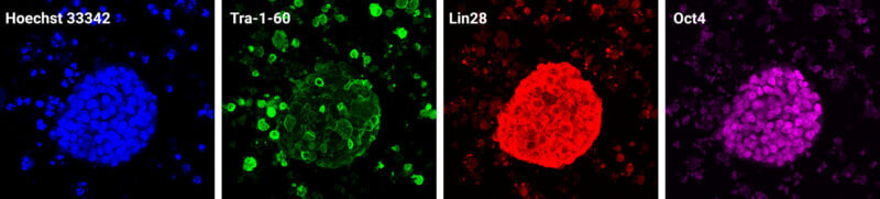
Figure 7: Indirect Immunofluorescence of Pluripotency Marker on IPSCs Grown in VitroGel® STEM Hydrogel
iPSCs (100k/well) were grown for 7 days in 24-well VitroPrime™ 3D Culture and Imaging Plate, fixed, and stained with pluripotency markers Tra-1-60, Lin28, and Oct4 using Alexa Fluor 488, 594, and 647 secondary antibodies. Images were captured using a Leica MICA confocal microscope. These results confirm that the RocketCell™ 3D iPSC Growth Kit, AOF provides a supportive 3D microenvironment for maintaining pluripotency.
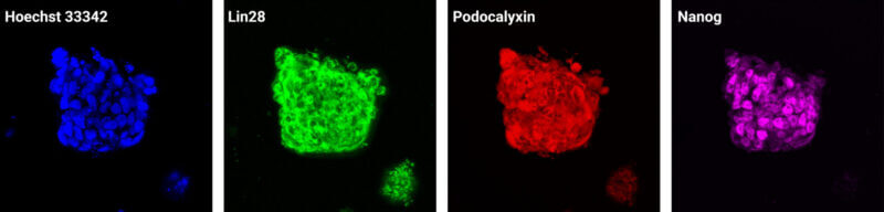
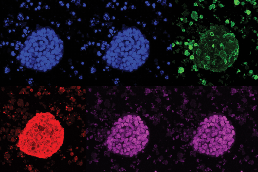
Figure 8: Direct Immunofluorescence Staining of IPSCs (HFF-1VL, TheWell Bioscience) Grown in VitroGel® STEM Hydrogel
iPSCs (100k/well) were grown for 7 days in 24-well VitroPrime™ 3D Culture and Imaging Plate before fixation and staining with directly labeled pluripotency markers Lin28 (Alexa 488), Podocalyxin (Alexa 594), and Nanog (Alexa 647). Images were captured using a Leica MICA confocal microscope. These results show that the RocketCell™ 3D iPSC Complete Growth Kit, AOF provides a supportive 3D environment for maintaining pluripotent stem cells, while the use of directly labeled antibodies enables faster and more efficient hydrogel-based sample processing.
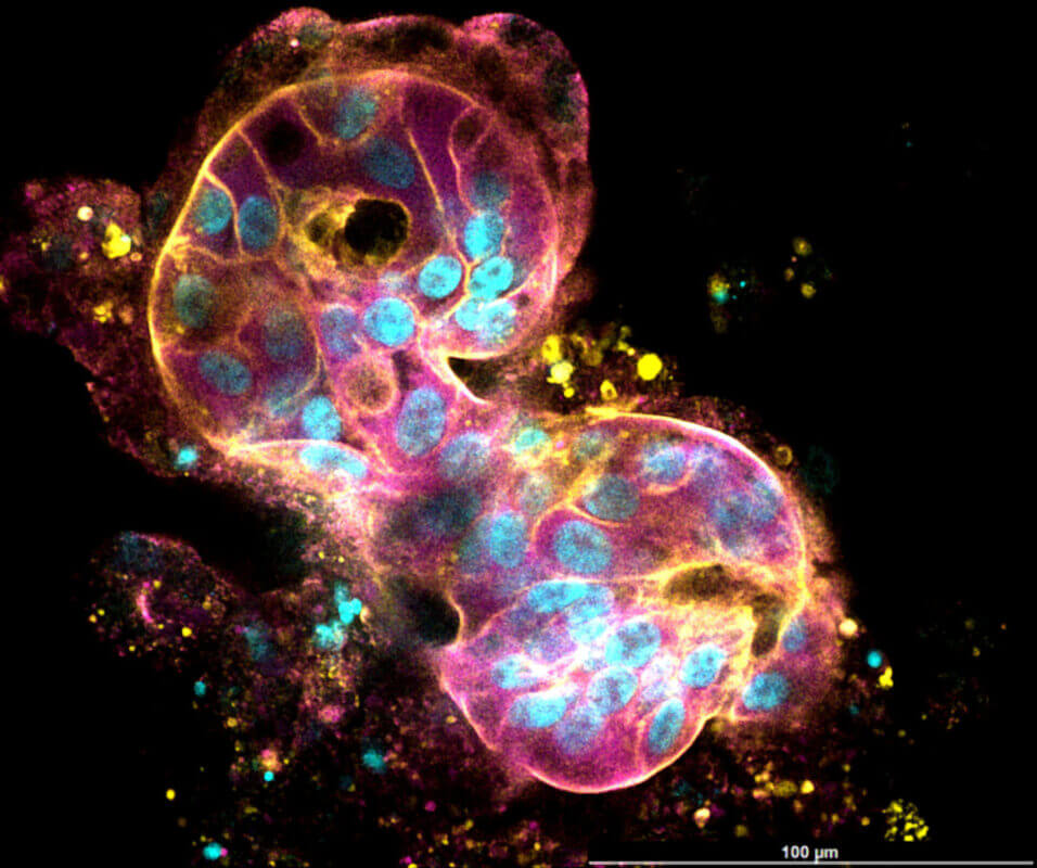
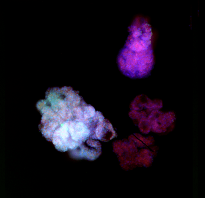
Figure 9. Use of VitroGel® Hydrogels to generate iPSC-derived instestinal organoids.
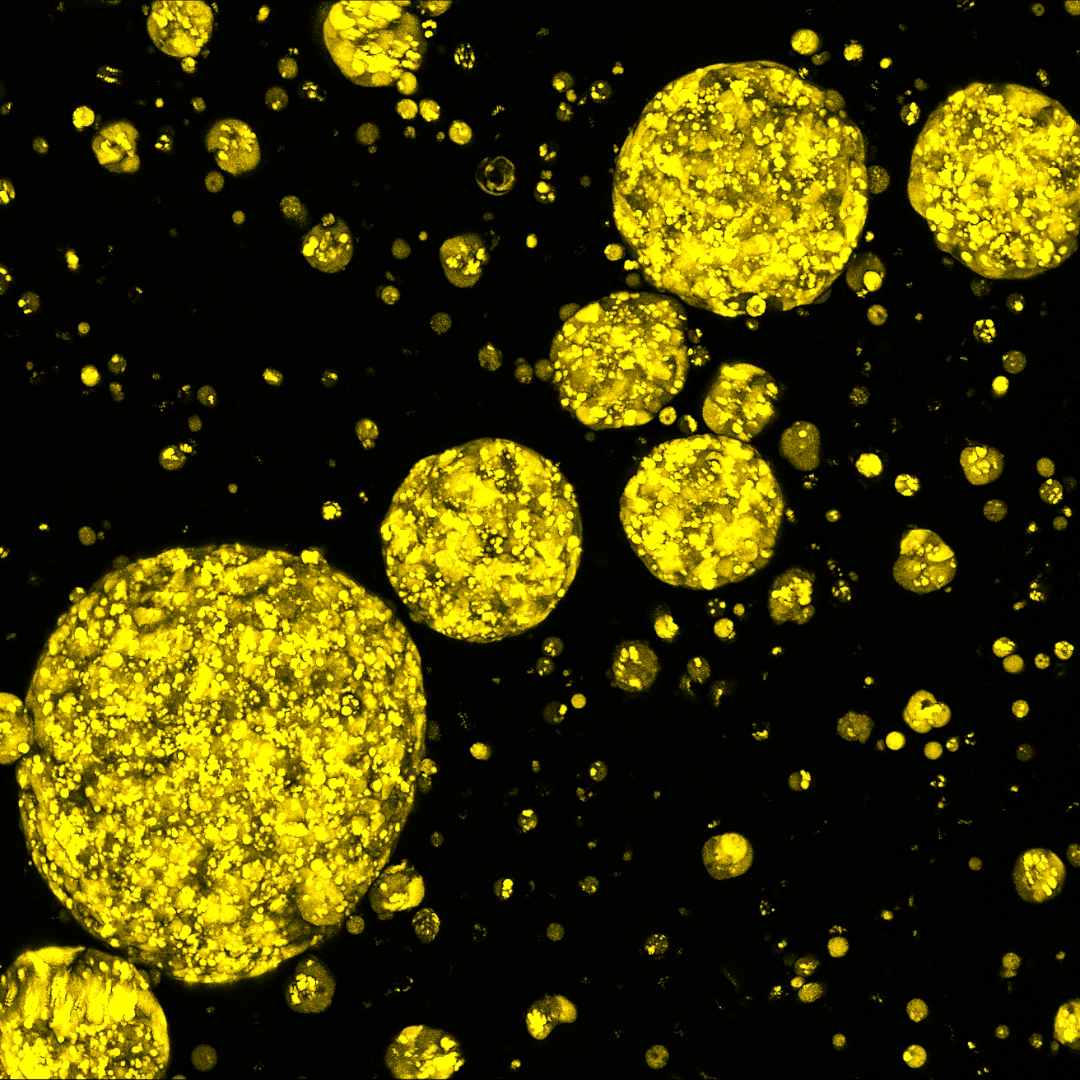
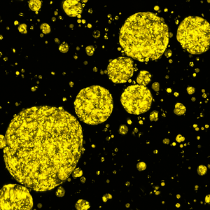
Representative image showing iPSC derived intestinal organoids cultured in VitroGel® ORGANOID. iPSC cells were cultured in VitroGel® STEM to generation of spheroids cells. The spheroids were then differentiated to intestinal organoids using CytoGrow™ growth factors. Yellow fluorescence: Phalloidin, Magenta: Villin protein, Blue staining (DAPI) represents cell nuclei.
Video Protocols
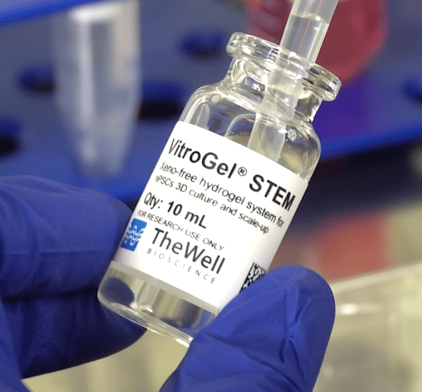
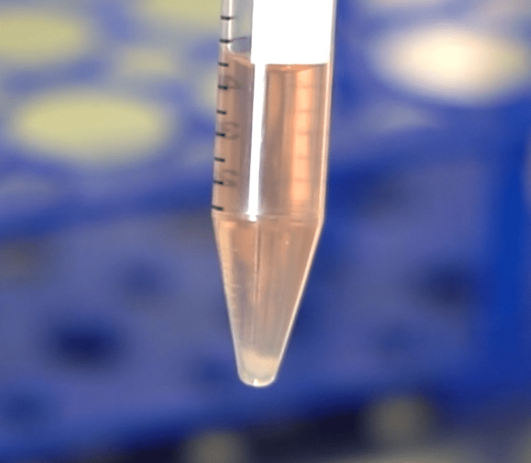
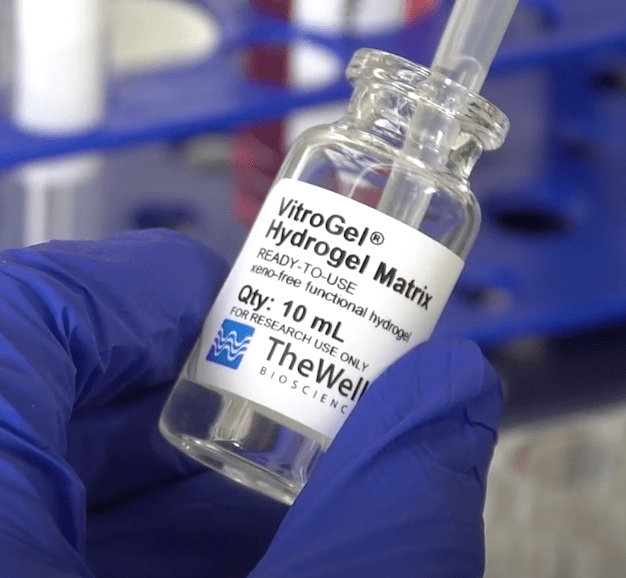

Enhance your stem cell culture with these products:
New
Cell Culture Media (Animal Origin-Free)
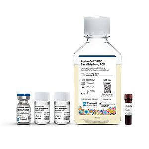
An all-in-one, animal origin-free kit with optimized matrix, medium, and reagents for 3D iPSC expansion.
New
Cell Culture Media (Animal Origin-Free)
Animal origin-free medium for 2D and 3D expansion of pluripotent stem cells.
New
Cell Culture Media (Animal Origin-Free)
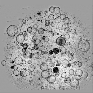
One Core Medium. Endless Organoid Possibilities. An animal origin-free medium that supports multiple organoid types - just add growth factors.
NEW
Cell Viability Enhancer
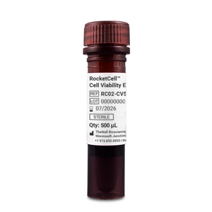
Maximize cell recovery and growth during passaging, thawing, and cryopreservation.
New
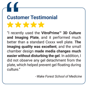
3D Culture Imaging Plates
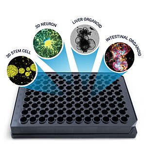
A premium cover-glass bottom plate for zero sample disruption 3D cell culture workflow: from cell seeding to high-resolution imaging.


3D Culture Vessels
Unique surface treated for superior hydrogel spreading, adherence, and uniform surface.
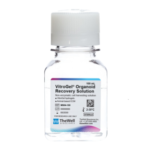
Cell Harvesting Solution
Chemically defined, non-enzymatic cell harvesting solution to recover cells/organoids from hydrogel or an animal-based ECM within 15 minutes.

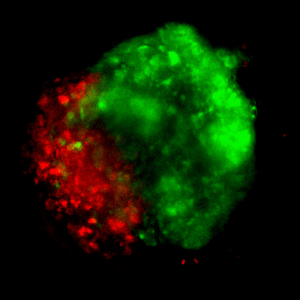
Downstream Cell Analysis
Fast (15 min), versatile, live/dead cell viability analysis for 3D and 2D cell culture.
References/Publications
| Size | 2 mL, 10 mL |
|---|
Related products
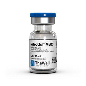
Hydrogels - Ready-To-Use
Ready-to-use, synthetic, chemically defined hydrogel system for mesenchymal stem cell 3D culture and scale-up
Hydrogels - Ready-To-Use
Ready-to-use, synthetic, chemically defined hydrogel system for 3D culture of HEK293 cells
NEW

Hydrogels - Ready-To-Use
Ready-to-use, synthetic, chemically defined hydrogel for neural stem cell and neuron cultures.
Hydrogels - Ready-To-Use
Ready-to-use, synthetic, chemically defined hydrogel system for organoid culture.
Hydrogels - Ready-To-Use
Ready-to-use, synthetic, chemically defined, biofunctional hydrogel system.
NEW
Hydrogels - Ready-To-Use
Ready-to-use, synthetic, chemically defined hydrogel that supports spheroid formation, tumoroid culture, and uniquely enables EMT biology Full xeno-free EMT kit available. Click here