

Premium low-attachment surface coating for 3D cultures
Excellent for spheroid generation and culture, organoid and tumoroid cultures, and suspension cultures.
New


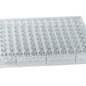

VitroPrime™ Ultra-Low Attachment Plate, U-Bottom, 96-Well
Premium U-bottom cell culture plate for 3D spheroids.

Premium VitroPrime™ Ultra-Low Attachment, U-Bottom, 96-well Plate
The VitroPrime™ Ultra-Low Attachment, U-bottom plate, 96-well Plate features a unique surface treatment that prevents cell adhesion, enabling spheroid formation and the creation of advanced 3D models, such as organoids and tumoroids. Designed with a uniform surface coating across wells, the VitroPrime™ Ultra-low Attachment plate ensures experimental consistency that enables drug screening and high-throughput applications.
Combined with the VitroGel® hydrogel system, researchers can examine spheroid invasion applications and complex tumoroid models.


Reduced cell adhesion
Unparalleled surface coating that prevents cell attachment, supporting consistent and rapid spheroid formation.

Uniform surface treatment
Homogeneous coating across wells that ensures experimental reproducibility and enables high-throughput and drug-screening applications.

Supports automated imaging systems
Works seamlessly with imaging platforms, including Incucyte from Sartorius, ImageXpress from Molecular Devices, and more.
Specifications
| Material | Polystyrene (GPPS) |
| Use | Spheroid formation and cultures, organoid and tumoroid cultures |
| Well Plate Type | 96-well |
| Well Profile | U-Bottom |
| Surface Type | Ultra-low adsorption |
| Sterile | Yes |
| Other Data | DNase/RNase-free, non-pyrogenic |
Protocols and Resources


Data and References
Spheroid Formation and Growth
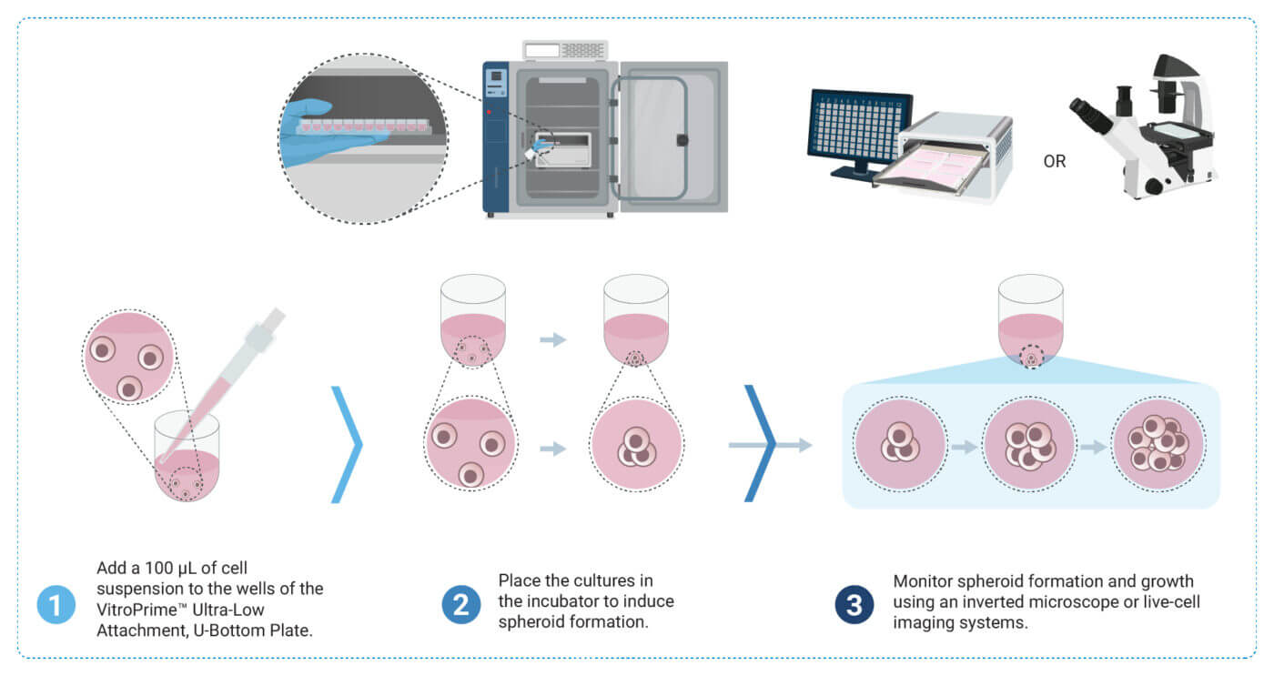
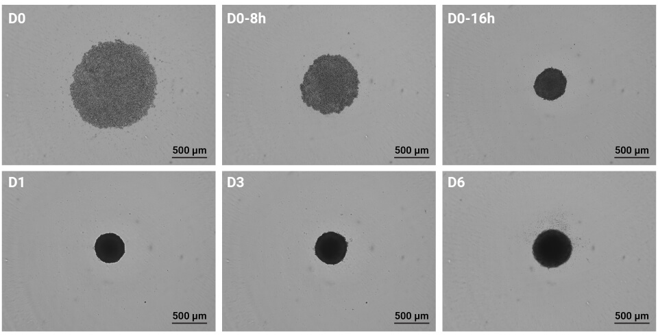
Figure 1. U87-MG GBM cells (2 x 10⁵ cells/mL) were resuspended in basal medium with supplement system. One hundred microliters (100 µL) of cell suspension were added to the VitroPrime™ Ultra-Low Attachment, U-Bottom, 96-Well Plate. Spheroid formation and growth were monitored with the Incucyte S3 live-cell analysis instrument on day 0 (0, 8, and 16 hours), and days 1, 3, and 6. The images were obtained at a 4X magnification.
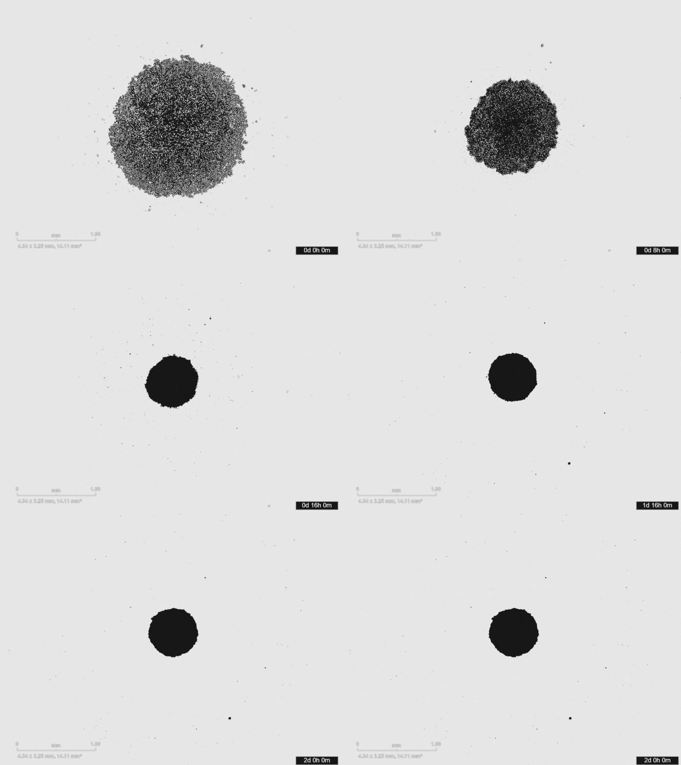
Figure 2. Time-lapse video depicting U87-MG glioblastoma spheroid formation in 48 hours. The video was obtained with the Incucyte S3 live-cell analysis instrument.
Comparison of Spheroid Formation between VitroPrime™ Ultra-Low Attachment, U-Bottom, 96-well Plate and 2 Commercially Available Ultra-Low Attachment Plates.
VitroPrime™ Ultra-Low Attachment,
U-Bottom, 96-well Plate
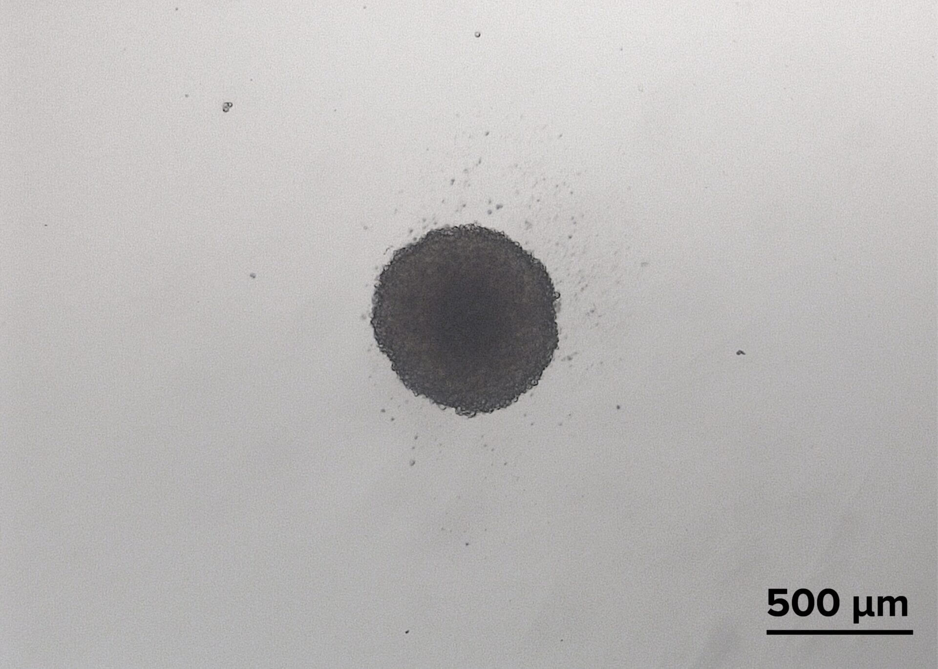
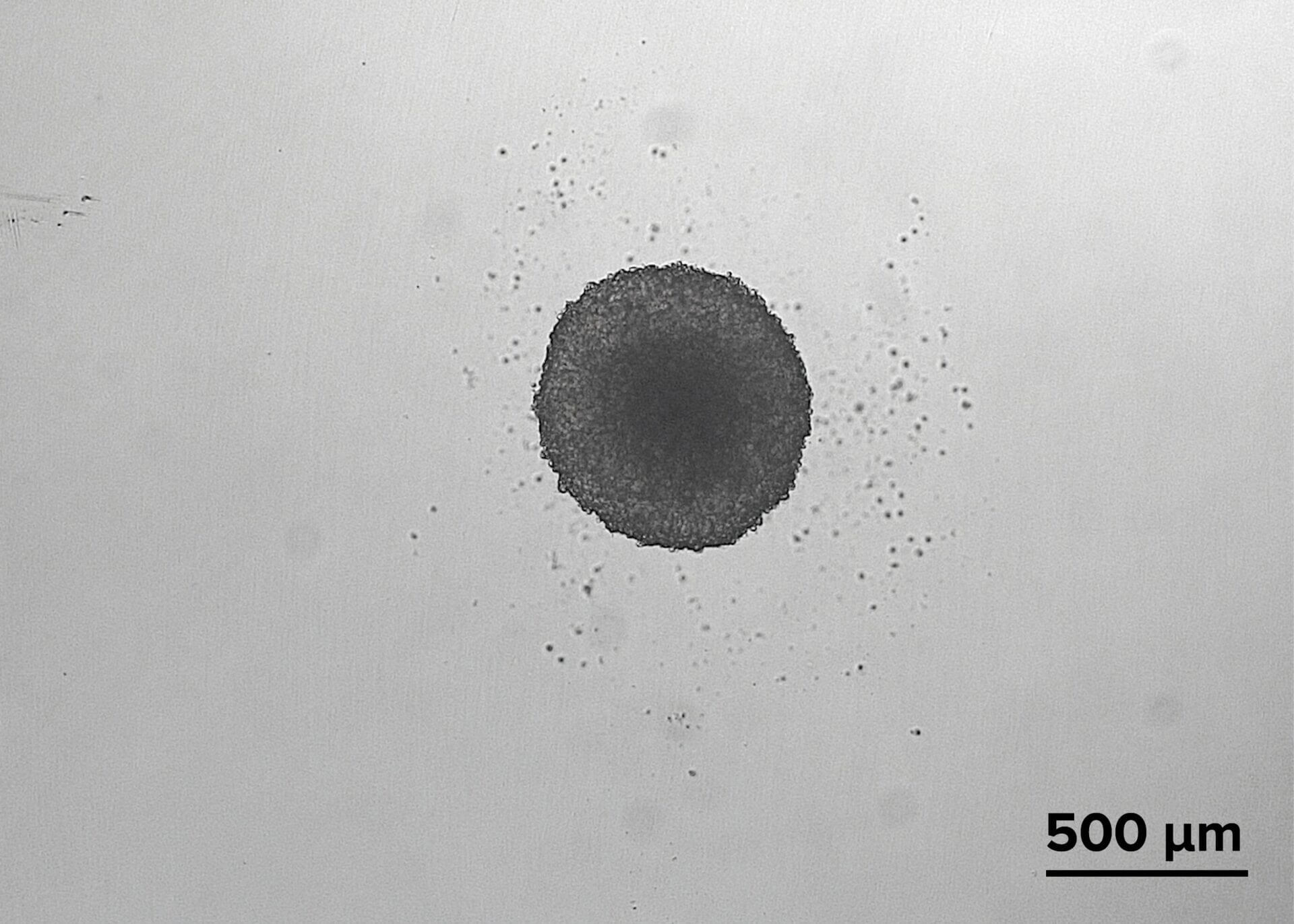
Premium
Ultra-Low Attachment Plate
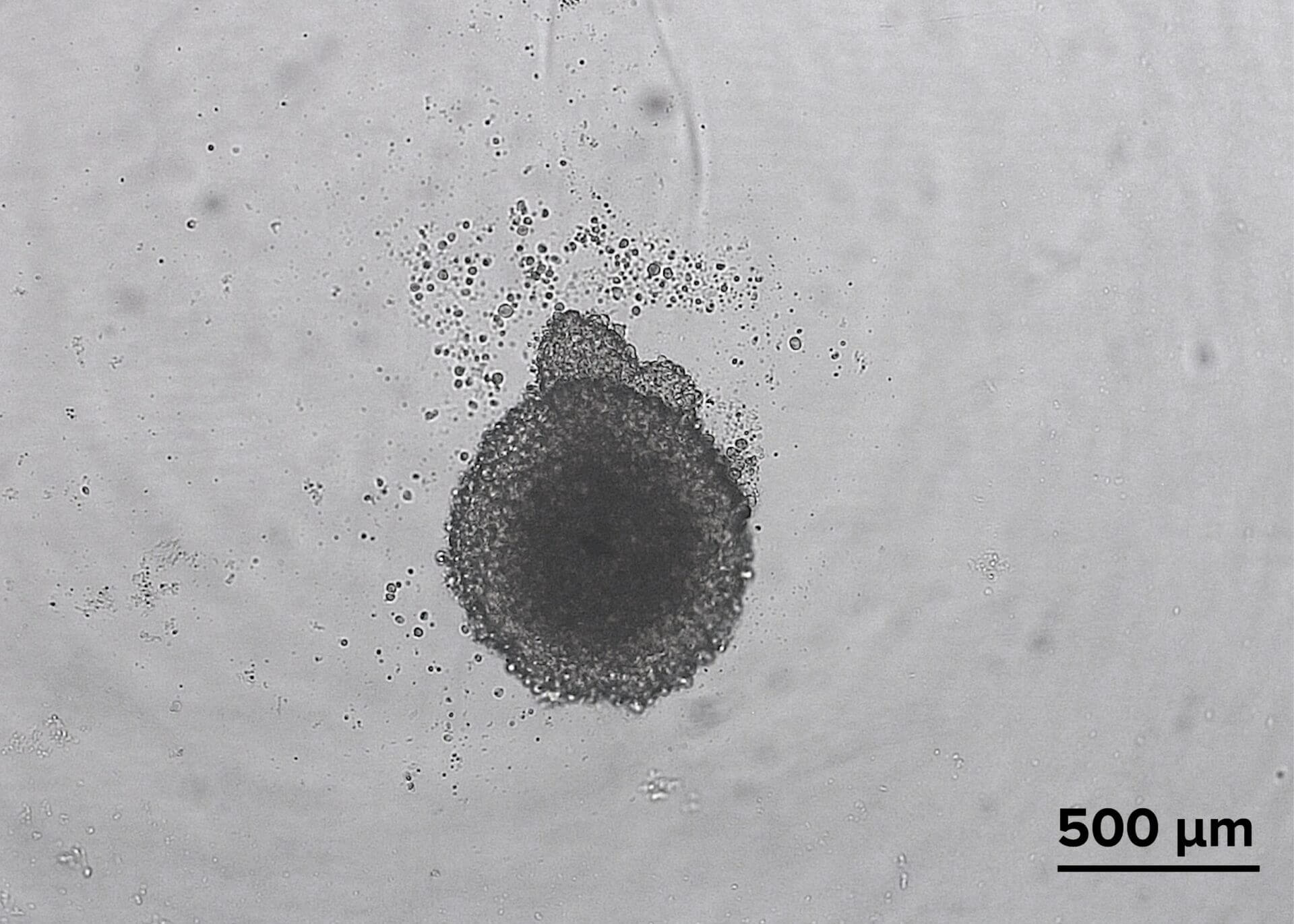
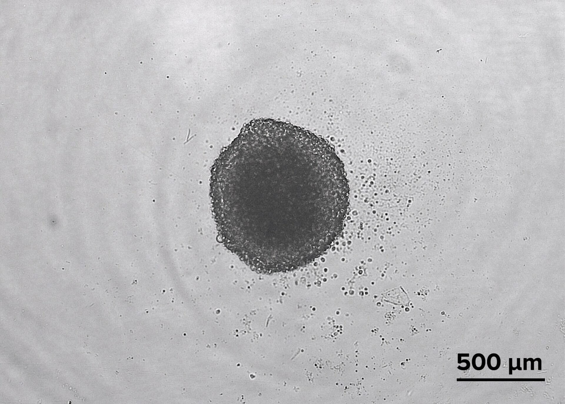
Standard
Ultra-Low Attachment Plate
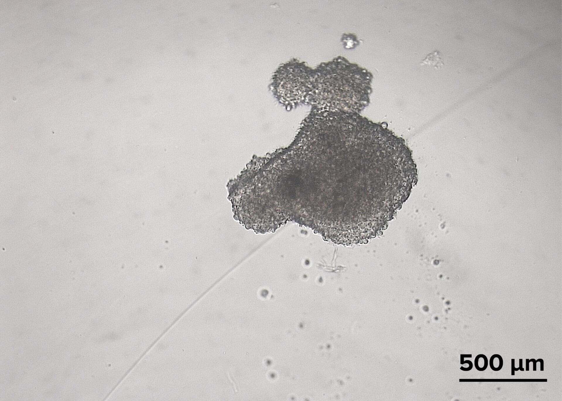
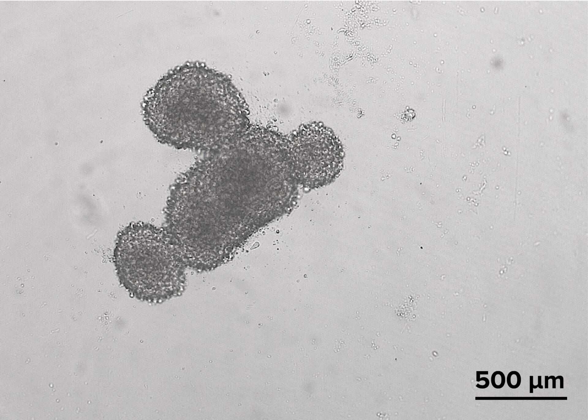
Figure 3. Comparison of spheroid formation between plates. The glioblastoma cells in the VitroPrime™ Ultra-Low Attachment, U-bottom, 96-well Plate formed a single spheroid, with no residual cells observed on the edges of the plate (Fig. 3, first column). However, cells in the commercially available plates failed to form round-shaped spheroids, which is crucial when performing spheroid invasion assays (Fig. 3, second and third columns).
Spheroid Invasion Assay
Achieve an easier and more consistent spheroid invasion when used with the VitroGel® hydrogel system.
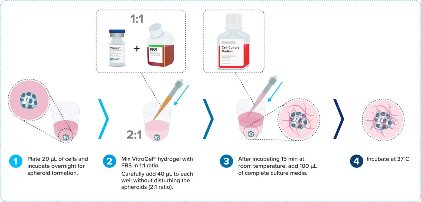
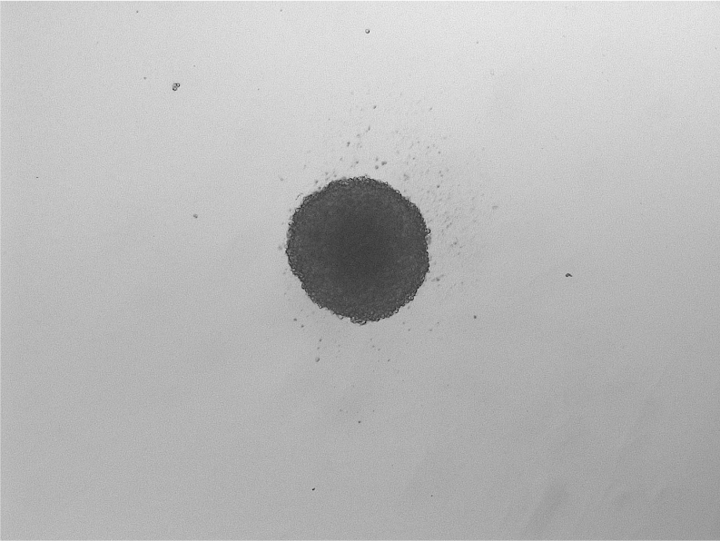
Spheroid Formation; No hydrogel (Day 0).
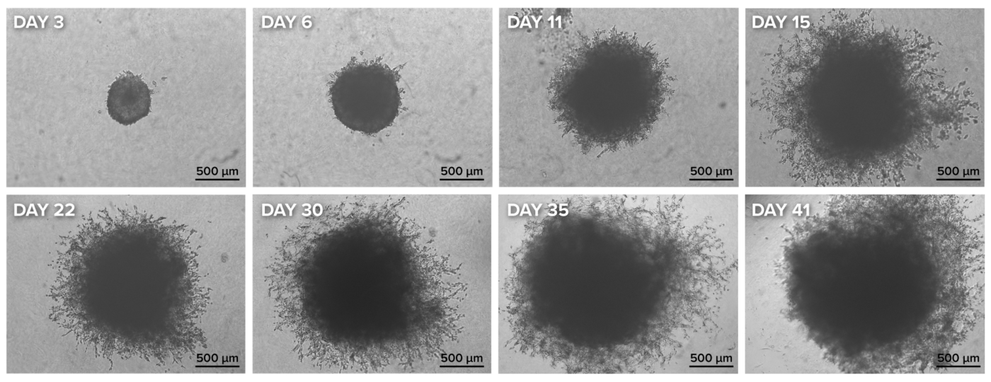
Figure 4. Spheroid invasion assay using the VitroPrime™ Ultra-Low Attachment Plate and VitroGel® Hydrogel Matrix
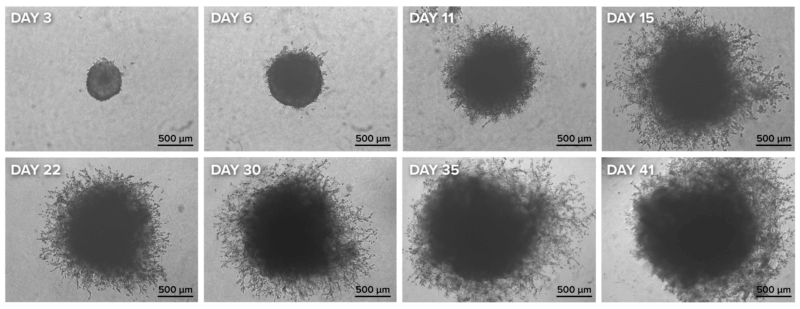
U87-MG glioblastoma cells were resuspended in basal medium with 10% fetal bovine serum. Twenty microliters (20 µL) of cell suspension were added to the VitroPrime™ Ultra-low Attachment Plate, U-bottom, 96-well plate. The cultures were incubated overnight at 37°C to allow spheroid formation. VitroGel® Hydrogel Matrix was combined with serum, and 40 µL of the mixture was added to the spheroid, followed by a 15-minute incubation at room temperature. The images were obtained with the Zeiss Microscope at a 2.5X magnification.
Epithelial to Mesenchymal Transition (EMT) Tumoroid
Create advanced 3D models with VitroGel® and VitroPrime™ Ultra-Low Attachment Plate.
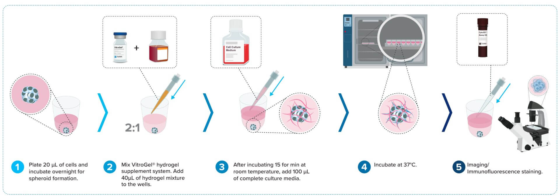
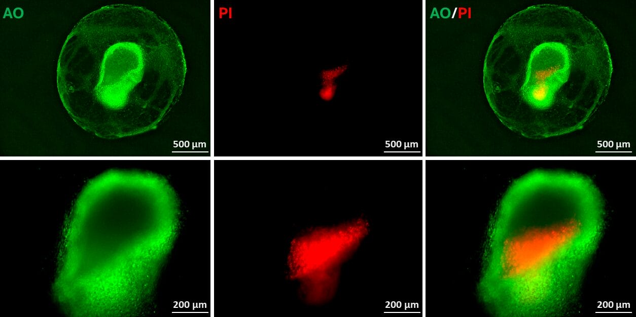
Figure 5. EMT GBM Tumoroid Viability
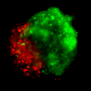
U87-MG GBM cells (1 x 10⁶ cells/mL) were resuspended in basal medium with the supplement system. Twenty microliters (20 µL) of cell suspension were added to the VitroPrime™ Ultra-low Attachment Plate, U-bottom, 96-well plate. The cultures were incubated overnight at 37 °C for spheroid formation. The hydrogel (40 µL) was added to the wells and incubated at room temperature for 15 minutes. A 100 µL of basal medium with supplements was added on top of the hydrogel. The cultures were incubated overnight, and the medium was changed every 2-3 days. After two weeks, the tumoroids were then subjected to Cyto3D® Live-Dead staining and carefully transferred to the VitroPrime™ Spread Attach 96-well plate. Acridine orange (AO) staining indicates the presence of live cells within the tumoroid as shown in green. Propidium iodide (PI; in red) stains for dead cells. Images were taken with the Keyence BZX microscope system at 4X (top) and 10X (bottom) magnifications.
Application Notes
Recommended Products
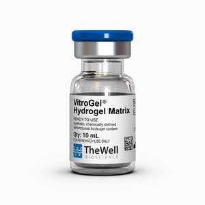
Hydrogels - Ready-To-Use
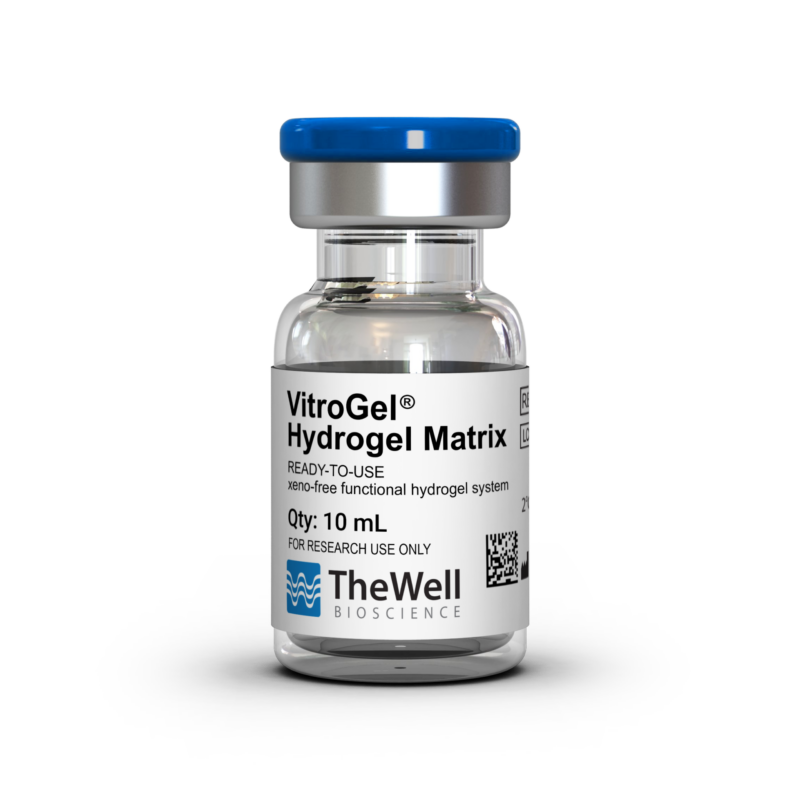
Ready-to-use, synthetic, chemically defined, biofunctional hydrogel system.

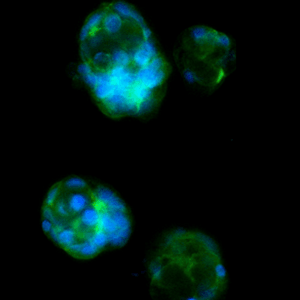
Downstream Cell Analysis
Fast (15 min), versatile, live/dead cell viability analysis for 3D and 2D cell culture.
| Quantity | 8 Packs, 8 Packs x 5 |
|---|
Related products
3D Culture Vessels
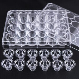
Transparent, PET, Sterile, 48 inserts (12/PK, 4PK/CS)
New

3D Culture Imaging Plates
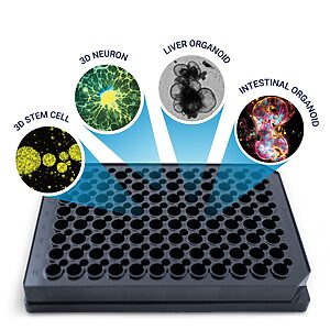
A premium cover-glass bottom plate for zero sample disruption 3D cell culture workflow: from cell seeding to high-resolution imaging.

3D Culture Vessels
Unique surface treated for superior hydrogel spreading, adherence, and uniform surface.