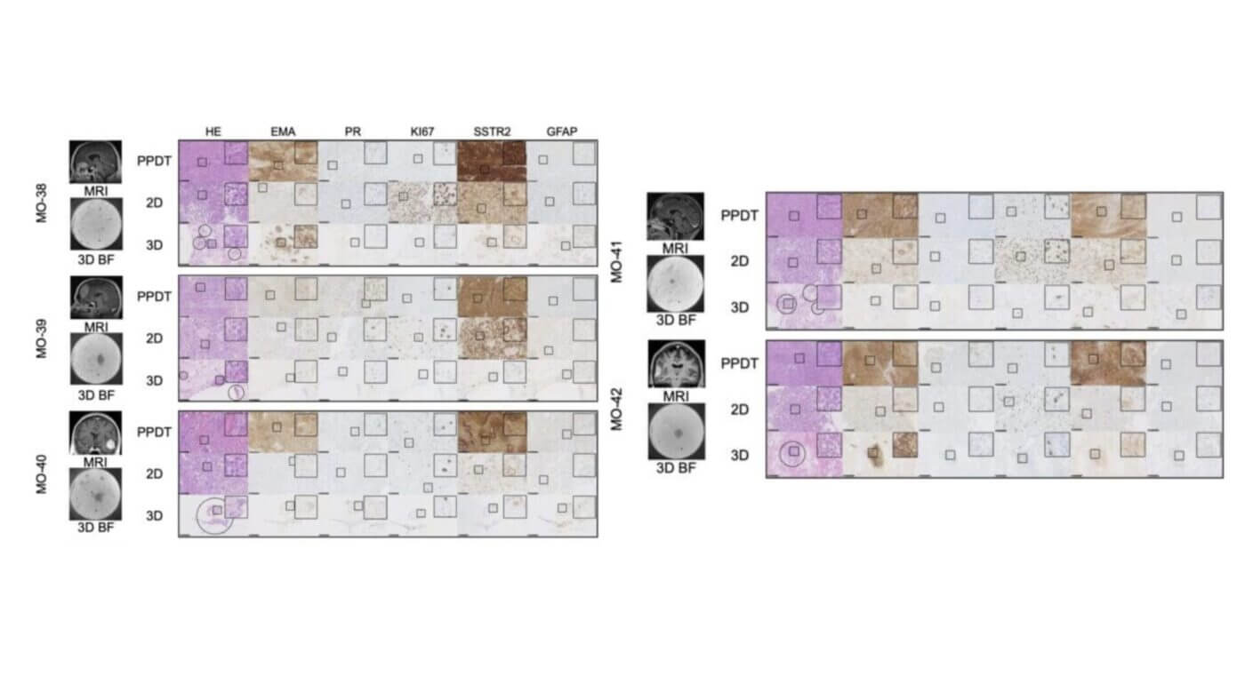
VitroGel® takes the fight to fibrosis—making cell invasion studies faster, clearer, and more reliable. Pulmonary fibrosis, especially when triggered by toxins such as paraquat, is a life-threatening condition driven by inflammation, epithelial-mesenchymal transition (EMT), and invasive cell behavior. To understand and halt this progression, researchers require a physiologically relevant in vitro system that can mimic […]
Category Archives: Research Highlights
This study leveraged VitroGel®’s hydrogel system to model the tumor microenvironment and uncover the role of TIMP1 in driving venous invasion in pancreatic cancer. Hydrogel: VitroGel® Hydrogel Matrix (VHM01) Pancreatic ductal adenocarcinoma (PDAC) is characterized by its aggressive nature and poor prognosis, often driven by early vascular invasion and metastasis. In this study, researchers focused […]
VitroGel® IKVAV was a key component that enabled stable vessel formation, proper cellular organization and functional BBB behavior in the 3D microfluidic model. Hydrogel: VitroGel® IKVAV (Cat. No: TWG007) The blood-brain barrier (BBB) presents a major challenge in neuro-oncology and CNS drug development due to its highly selective nature, which restricts the passage of most […]
VitroGel® Hydrogel Matrix enables the controlled delivery of bioactive Qu-SeNPs, accelerating bone defect healing and enhancing therapeutic potential for regenerative medicine. Hydrogel: VitroGel® Hydrogel Matrix (Cat. No: VHM01) The development of biomimetic hydrogels has significantly advanced the field of regenerative medicine by providing an extracellular matrix-like environment that supports cell survival and function. In bone […]
VitroGel® supports 3D spheroid models for evaluating basroparib’s efficacy in overcoming MEK inhibitor resistance in KRAS-mutated colorectal cancer. Hydrogel:VitroGel® RGD (Cat. No: TWG003) The study on basroparib’s role in overcoming MEK inhibitor resistance in KRAS-mutated colorectal cancer (CRC) highlights the importance of innovative in vitro models that accurately recapitulate tumor microenvironments. Using VitroGel®, a versatile, […]
VitroINK® provided a xeno-free, structurally supportive bioink that enabled the precise 3D bioprinting of a functional testis model, facilitating cell viability, maturation, and in vitro spermatogenesis research. Fertility preservation following pediatric cancer therapy has become a critical area of research, as over 80% of childhood cancer survivors reach adulthood, yet up to 66% of male […]
VitroGel® 3D provided a scaffold-free yet supportive matrix that stabilized adipose organoids for imaging and analysis, enhancing the reliability of in vitro obesity research. Obesity and metabolic diseases remain urgent global health challenges, yet studying human adipose tissue in vitro is complicated by the limitations of 2D cultures and scaffold-based models. Traditional methods often fail […]
Biomarker Cyto3D® Live-Dead Assay Kit (Cat. No: BM01) The study explores the application of tumor organoid technology in borderline ovarian tumors (BOT), a category of tumors with limited treatment options and resistance to conventional chemotherapy. Researchers established patient-derived organoids (PDOs) to model BOT and evaluate potential therapeutic compounds, particularly Bractoppin, a BRCA1 carboxy-terminal domain (BRCT) […]
Hydrogel: VitroGel® ORGANOID 3 (Cat. No: VHM04-3) Pancreatic ductal adenocarcinoma (PDAC) is one of the deadliest cancers, with limited treatment options and poor patient survival rates. A major challenge in treating PDAC is the tumor’s ability to create a protective barrier of mucins, which shields cancer cells from immune attacks and reduces the effectiveness of […]
BiomarkerCyto3D® Live-Dead Assay Kit (Cat. No: BM01) Cancer research has long been challenged by the complexity of tumor-immune interactions. Traditional 2D cell cultures and animal models often fail to capture the intricate tumor microenvironment (TME), limiting their relevance to human biology. Patient-derived precision-cut tumor slices (PCTS) offer a promising solution by preserving the tumor’s architecture […]


