Poster Presentations
The Growth and Neuronal Differentiation of Human Induced Pluripotent Stem Cells in Xeno-Free Functional Synthetic Hydrogel
Abstract #6425 at SfN – Neuroscience 2025
The Growth and Neuronal Differentiation of Human Induced Pluripotent Stem Cells in Xeno-Free Functional Synthetic Hydrogel
PSTR100.17/A17
Abstract:
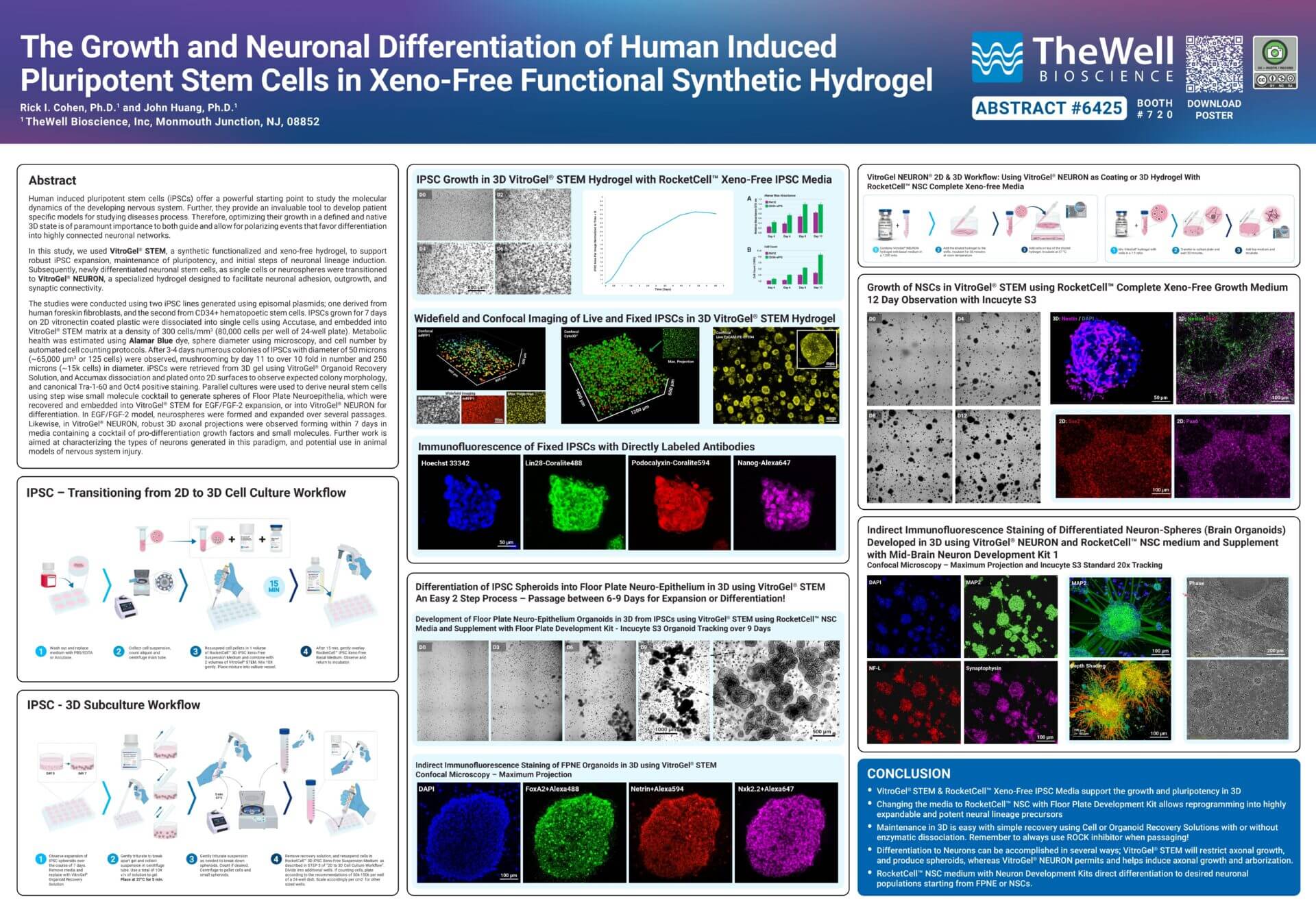
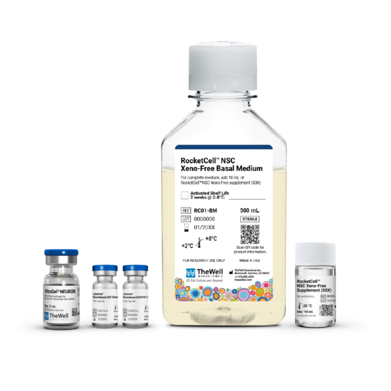
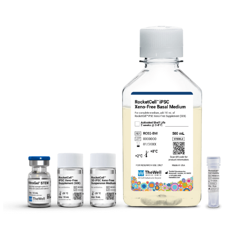
Human induced pluripotent stem cells (iPSCs) offer a powerful starting point to study the molecular dynamics of the developing nervous system. Further, they provide an invaluable tool for developing patient-specific models to study the disease process. Therefore, optimizing their growth in a defined, native 3D state is of paramount importance to both guide and enable polarizing events that favor differentiation into highly connected neuronal networks. In this study, we used VitroGel STEM®, a synthetic, functionalized, xeno-free hydrogel, to support robust iPSC expansion, maintenance of pluripotency, and the initial steps of neuronal lineage induction. Subsequently, newly differentiated neuronal stem cells, as single cells or neurospheres, were transitioned to VitroGel® NEURON, a specialized hydrogel designed to facilitate neuronal adhesion, outgrowth, and synaptic connectivity.
The studies were conducted using two iPSC lines generated using episomal plasmids: one derived from human foreskin fibroblasts and the second from CD34+ hematopoietic stem cells. IPSCs grown for 7 days on 2D vitronectin-coated plastic were dissociated into single cells using Accutase and embedded into VitroGel® STEM matrix at a density of 300 cells/mm3 (80,000 cells per well in a 24-well plate). Metabolic health was estimated using Alamar Blue dye, sphere diameter was measured by microscopy, and cell number was determined using automated cell counting protocols. After 3-4 days, numerous colonies of IPSCs with a diameter of 50 microns (~65,000 um3 or 125 cells) were observed, mushrooming by day 11 to over 10-fold in number and 250 microns (~15k cells) in diameter.
iPSCs were retrieved from 3D gel using VitroGel® Organoid Recovery Solution, and Accumax dissociation and plated onto 2D surfaces to observe expected colony morphology, and canonical Tra-1-60 and Oct4 positive staining. Parallel cultures were used to derive neural stem cells using a stepwise small-molecule cocktail to generate spheres of Floor Plate Neuroepithelia, which were recovered and embedded in VitroGel® STEM for EGF/FGF-2 expansion or in VitroGel® NEURON for differentiation. In the EGF/FGF-2 model, neurospheres were formed and expanded over several passages. Likewise, in VitroGel® NEURON, robust 3D axonal projections were observed forming within 7 days in media containing a cocktail of pro-differentiation growth factors and small molecules. Further work aims to characterize the types of neurons generated in this paradigm and their potential use in animal models of nervous system injury.
Please complete the form below to download the poster.
Related Products
Discover how VitroGel® can support your cell culture research
Explore our updated resources for a concise overview of VitroGel®’s key features and application areas.
Comparison of VitroGel® vs. Animal-based ECM
Discover the 20+ advantages of VitroGel® over animal-based ECM with this comprehensive comparison on key features, operation, application, and storage conditions.
Learn More
What is VitroGel® | Overview Video
Learn why VitroGel® is the leading animal-free hydrogel for 3D cell culture.
Learn More