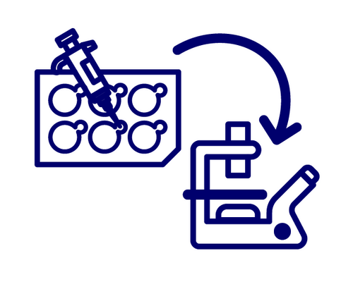
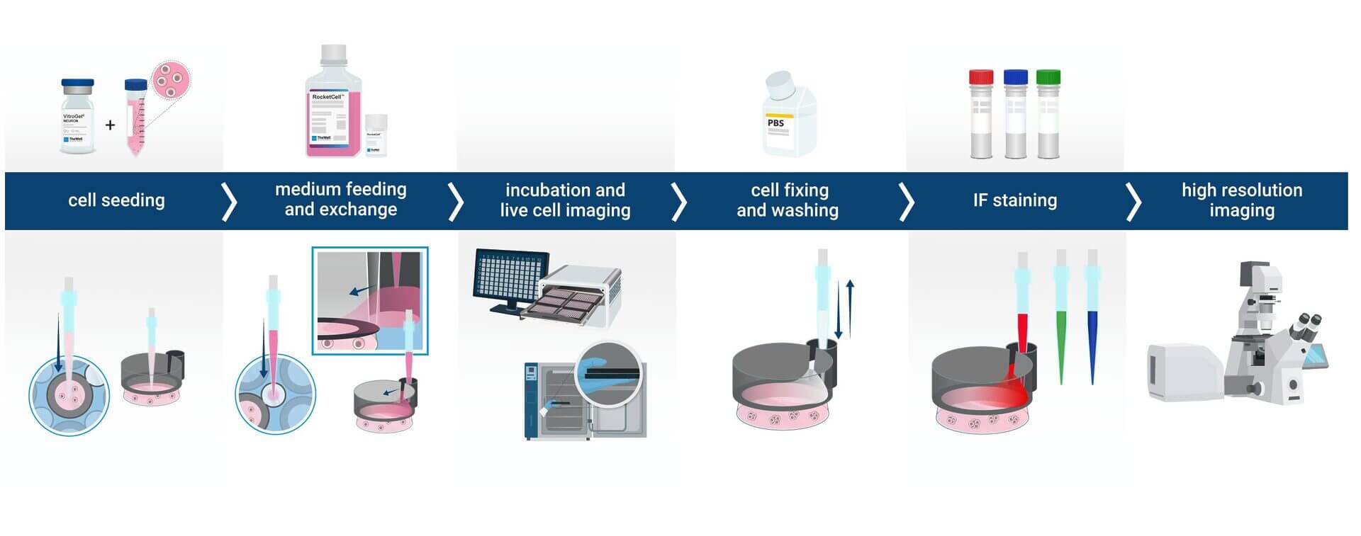
End-to-End Workflow Integration
Supports a complete “in-plate” workflow: embedding, polymerization, long-term culture, fixation, and IF staining. No sample transfer means no sample loss.
New
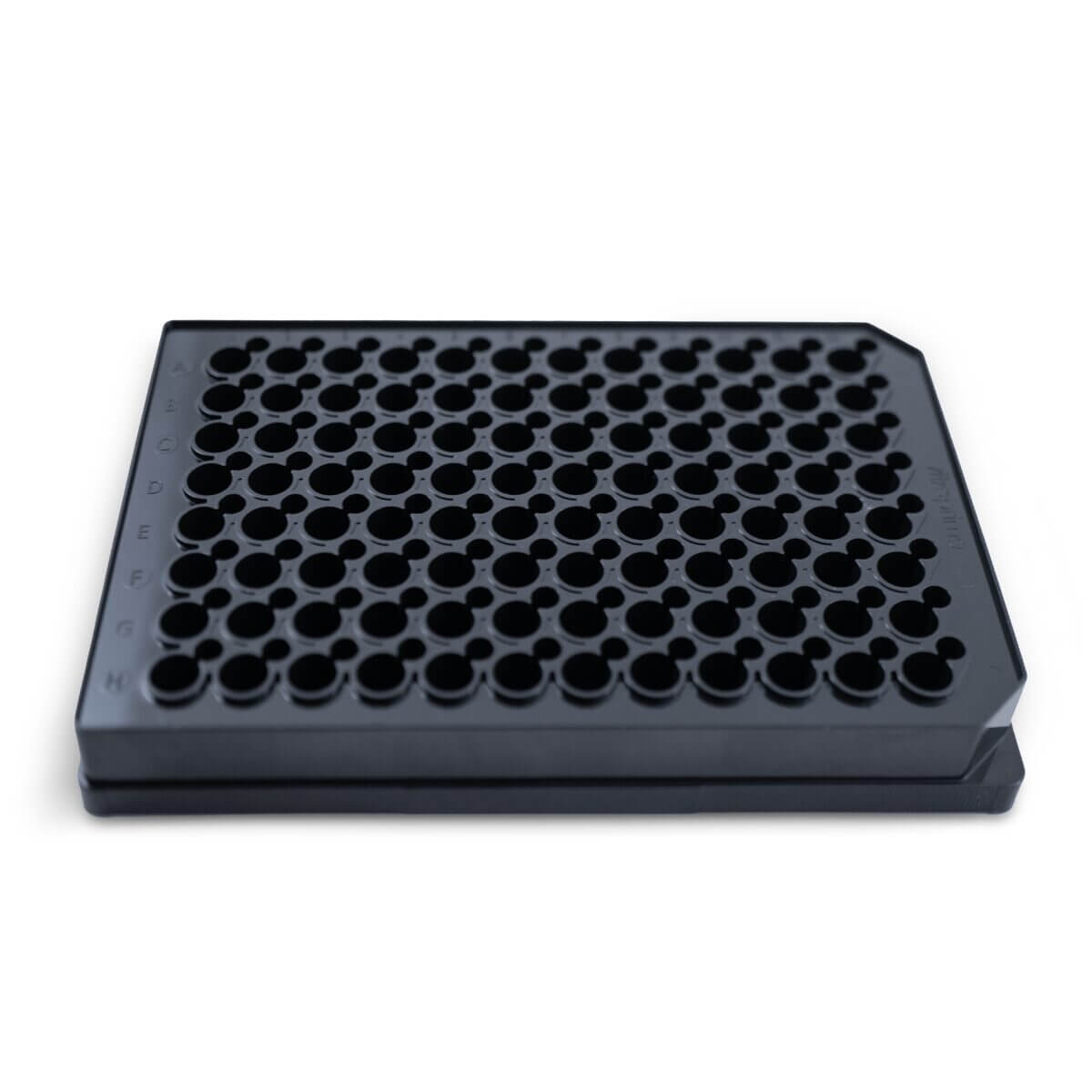

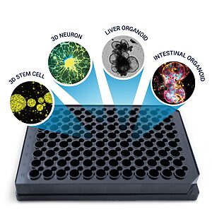

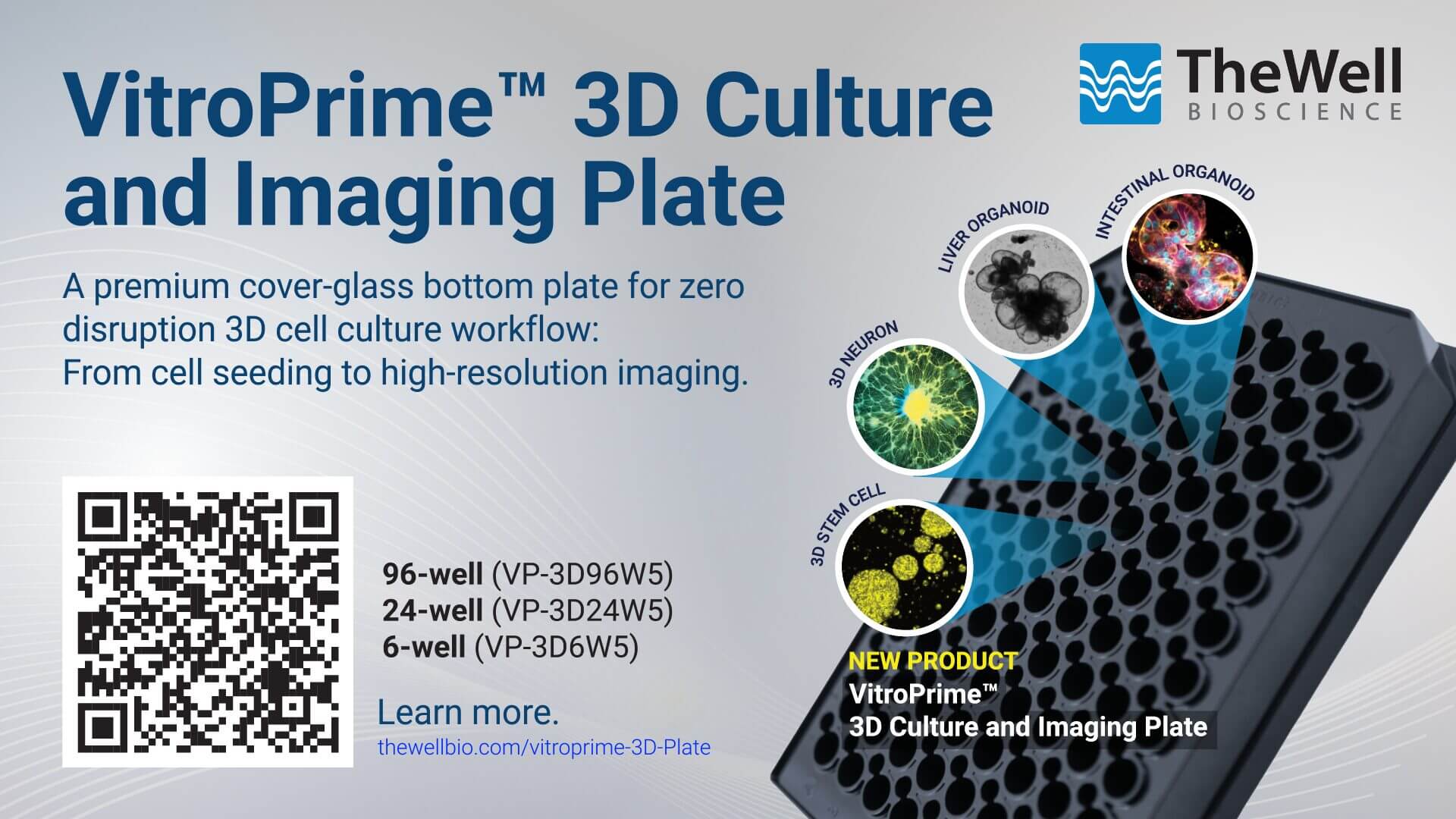
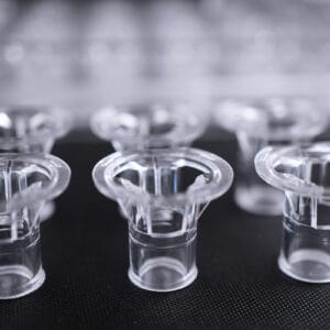
VitroPrime™ 3D Culture and Imaging Plate
A premium cover-glass bottom plate for zero disruption 3D cell culture workflow: From cell seeding to high-resolution imaging.
 3D Culture and Imaging Plate
Premium 3D cell culture plate featuring a unique media-exchange channel and “sample-locking” technology, combined with an ultra-clear, premium cover-glass bottom, enabling a zero-disruption 3D cell culture workflow.
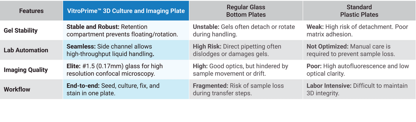
Using standard cell culture plates for 3D cell culture introduces several challenges:
- Gel Floating and rotation make imaging and tracking difficult.
- Precious samples are at risk of being lost during media changes.
- Multiple vessel transfers during washing and staining increase handling steps.
- Repeated handling can disrupt or damage delicate 3D structures.
- Limited compatibility with automated workflows.
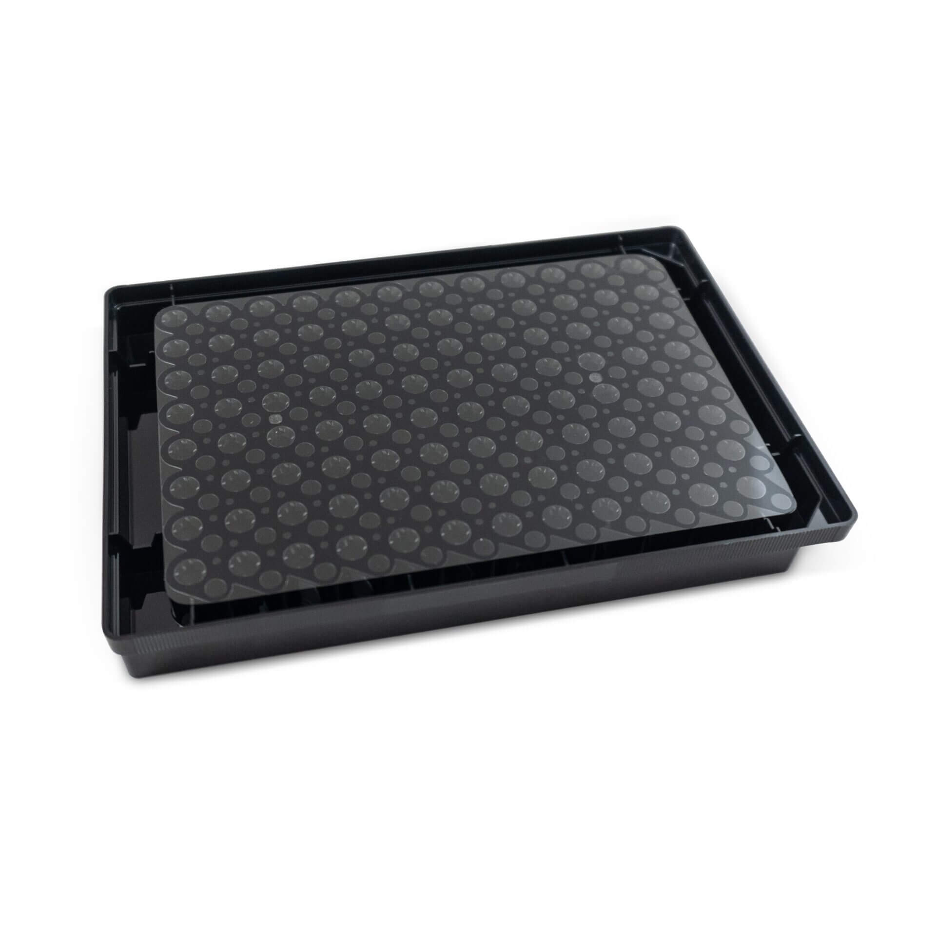
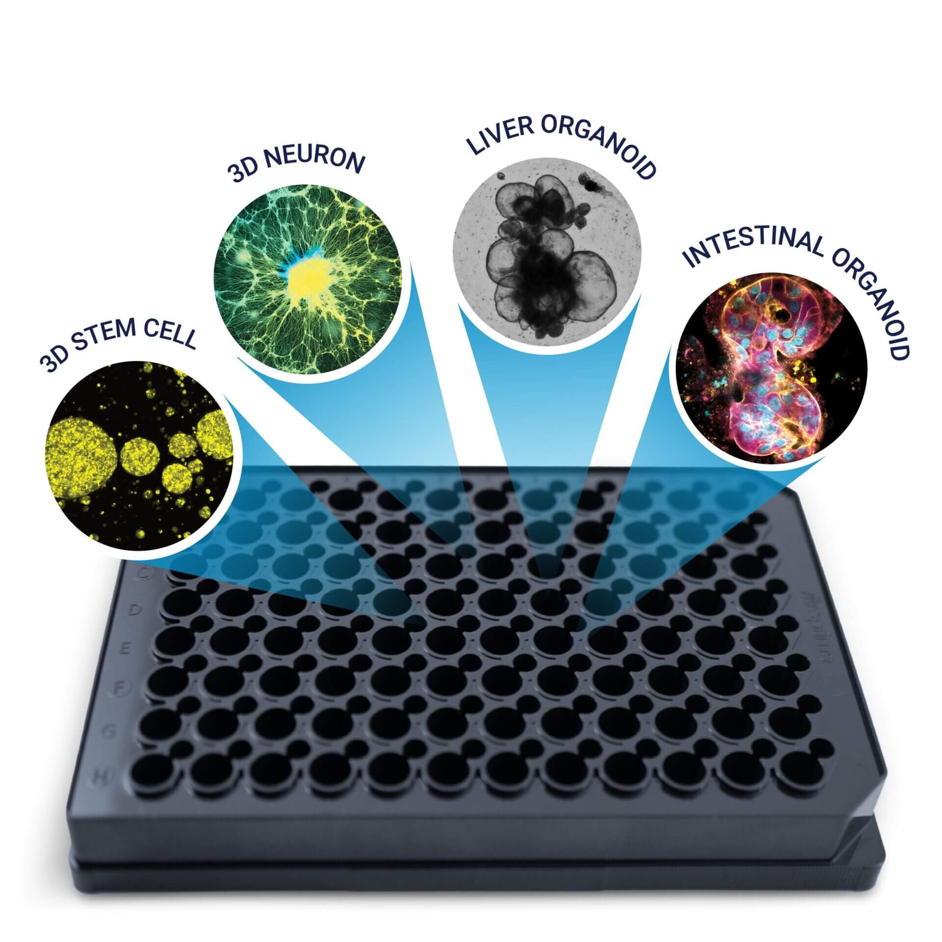
VitroPrime™ 3D Culture and Imaging Plates are specifically designed to solve the common challenges of 3D hydrogel culture. Engineered with a built-in retention compartment keeps the gel firmly in place, preventing floating and rotation for stable imaging and accurate tracking. By allowing culture, media changes, washing, staining, and imaging to be performed in a single vessel, VitroPrime™ 3D Culture and Imaging Plate reduces sample loss, protects delicate 3D structures, simplifies handling, and supports automation-friendly workflows.

Disturbance-Free Medium Exchange
A separate, dedicated channel allows for the smooth diffusion of nutrients and reagents without the risk of aspirating or disrupting the 3D hydrogel structure.

Anti-Floating & Anti-Rotation Technology
The integrated hydrogel retention compartment ensures the matrix stays securely anchored. No more lost samples during medium changes or “drifting” objects during time-lapse imaging.

Elite Optical Clarity
The premium #1.5 cover-glass bottom provides maximum light transmission and minimal auto-fluorescence, optimized for high-resolution confocal and immersion-objective imaging.

Automation Ready
Fully compatible with automated liquid-handling systems and high-content imaging (HCI) platforms.
Redefining the 3D Cell Culture Workflow (In-Plate Workflow)
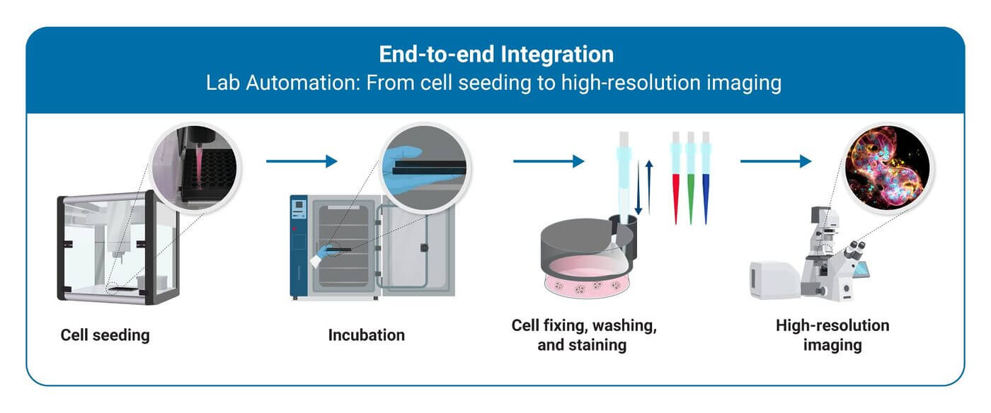
VitroPrime™ 3D Culture and Imaging Plates are premium-tier vessels engineered to streamline end-to-end workflows for hydrogel-based 3D culture. By integrating every step from cell embedding and matrix polymerization to long-term culture, downstream immunofluorescence (IF) staining, and high-resolution imaging into a single high-performance platform, VitroPrime™ 3D Culture and Imaging Plate minimizes sample loss and ensures consistent, reproducible results.
Mechanical Anchoring for Precision Tracking
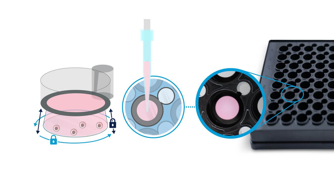
Unlike standard plates, where gels can detach, the VitroPrime™ 3D Culture and Imaging Plate securely anchors the gel with a built-in retention compartment. This prevents gel floating or rotation, keeping organoids stable for reliable live-cell imaging and long-term tracking.
Zero-Disruption Automation and Medium Exchange
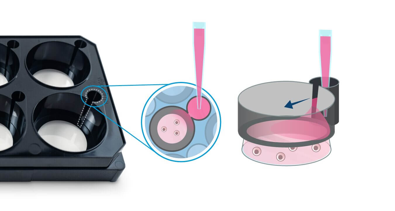
Each well includes a lateral medium-exchange channel that protects the delicate 3D environment. Nutrients and reagents diffuse gently without disturbing the hydrogel, enabling automated, high-throughput media changes, fixation, and staining—without risking damage to the 3D matrix.
Elite Optical Performance
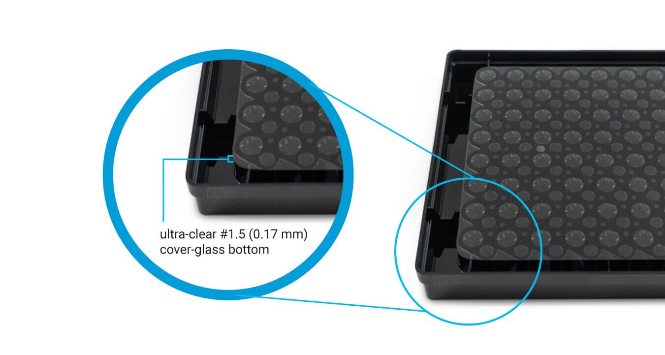
The VitroPrime™ 3D Culture and Imaging Plate uses an ultra-clear #1.5 (0.17 mm) cover-glass bottom for superior imaging. It maximizes light transmission, minimizes auto-fluorescence, and delivers high-resolution images. Ideal for confocal microscopy, immersion objectives, and high-content screening.
Specifications
Material Polstyrene molding and #1.5 (0.17 mm) Ultra-clear glass bottom
Packaging 5 packs/case
Well Plate Type 6-well, 24-well, 96-well
Well Profile • Flat bottom
• Optimized compartment for hydrogel retention
• Dedicated lateral medium-exchange channel
Sterilization Gamma Radiation
Other Data DNase/RNase-free, non-pyrogenic
Shelf Life 15 months
Product Documentation
![]() VitroPrime™ 3D Culture and Imaging Plate – Sale Sheet
VitroPrime™ 3D Culture and Imaging Plate – Sale Sheet
![]() Frequently Asked Questions
Frequently Asked Questions
Data and References
End-to-End Workflow Integration & Disturbance-Free Medium Exchange

Anti-Floating and Anti- Rotation Technology Case 1:
Live Imaging of mRFP1-labeled iPSCs in a 96-well VitroPrime™ 3D Culture and Imaging Plate
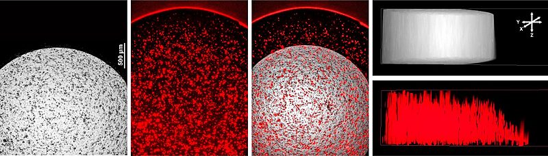
Fig 1. Growth of mRFP-labeled IPSCs in 96-well VitroPrime™ 3D Culture and Imaging Plate
IPSCs were harvested from 2D sources using Accutase and plated into the 96-well VitroPrime™ 3D Culture and Imaging Plate, as recommended, at a volume of 20 uL per well containing 20,000 cells/well. Cells were grown for 5 days prior to imaging using a Keyence BZX imaging system with z-stacking through the entire 1.5 mm hydrogel thickness. The brightfield (transmitted) and mRFP (reflected) images were compiled using a full-focus algorithm and displayed along the Z-axis to help visualize the shape of the light used in both imaging techniques. This data shows that cells under the angled black retaining rim thrive and grow as if in the center of the well.
Anti-Floating and Anti- Rotation Technology Case 2:
Mechanical Anchoring Enables Precision Tracking andPrevents Gel Floating and Rotation
in Liver Organoids
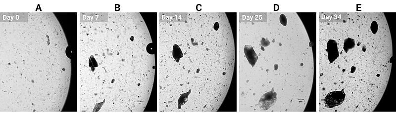
Fig 2. VitroPrime™ 3D Culture and Imaging Plate prevents gel floating and rotation of liver organoids.
(A-E) Images indicate liver organoid growth over 30 days at the same location, cultured in VitroGel® ORGANOID in a 24-well VitroPrime™ 3D Culture and Imaging Plate.

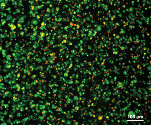
Elite Optical Clarity Case 1:
Live-Dead Staining of iPSCs Grown in VitroGel® STEM ( No wash protocol)
A
B
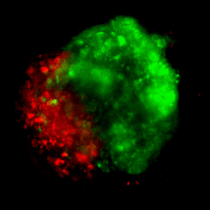
Fig 3. Cyto3D® Live-Dead Assay for Monitoring the Health of IPSC (HFF-1VL, TheWell Bioscience) Grown in RocketCell™ 3D iPSC Xeno-Free Complete Growth Kit.
IPSCs (100k/well) from 2D sources were plated in a 24-well, VitroPrime™ 3D Culture and Imaging Plate. After 7 days, the medium was removed and replaced with media containing a 1:50 dilution of the Cyto3D® Live-Dead Assay solution. After 30 min, the well was imaged on a Leica MICA confocal microscope (A, B). The rendered image (B) is 600 microns deep, with a tiled field approximately 1.4 mm x 1.1 mm. The data also demonstrate that the Cyto3D® protocol does not require any wash steps and can expedite imaging protocols used to assess the vitality of live cultures.
Elite Optical Clarity Case 2:
Live EpCAM Cell Surface Immunostaining of IPSCs Grown in VitroGel® STEM
(Confocal Microscopy)
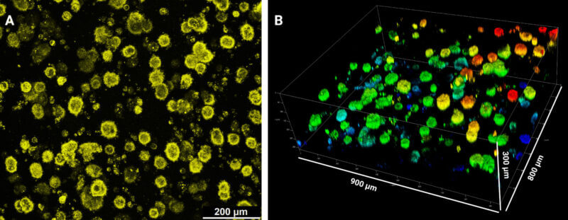
Fig 4. Live Cell Surface Immunofluorescent Staining of Human IPSCs (HFF-1VL, TheWell Bioscience) Grown in RocketCell™ 3D iPSC Xeno-Free Complete Growth Kit.
IPSCs (100k/well) from 2D sources were plated in a 24-well VitroPrime™ 3D Culture and Imaging Plate. After 7 days, the medium was removed and replaced with media containing a diluted anti-EPCAM-PE-Alexa594-labeled antibody. The mixture was incubated for 1 hour at 37 °C, and then the well was rinsed three times with 5 min incubation with 0.5 mL of Growth Media. The well was imaged on a Leica MICA confocal microscope (A). The rendered image (B) is 300 microns deep, with a field approximately 800 x 900 microns.
Elite Optical Clarity Case 3:
Indirect Immunofluorescence staining of IPSCs Grown in VitroGel® STEM
Pluripotency is maintained by RocketCell™ iPSC Xeno-Free Growth Medium
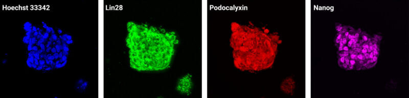
Fig 5. Direct Immunofluorescence Staining of IPSCs (HFF-1VL, TheWell Bioscience) Grown in RocketCell™ 3D iPSC Xeno-Free Complete Growth Kit.
iPSCs (100k/well) were grown for 7 days in a 24-well VitroPrime™ 3D Culture and Imaging Plate before fixation and staining with directly labeled pluripotency markers Lin28 (Alexa 488), Podocalyxin (Alexa 594), and Nanog (Alexa 647). Images were captured using a Leica MICA confocal microscope. These results show that the RocketCell™ 3D iPSC Xeno-Free Complete Growth Kit provides a supportive 3D environment for maintaining pluripotent stem cells, while the use of directly labeled antibodies enables faster and more efficient hydrogel-based sample processing.
Elite Optical Clarity Case 4:
Immunofluorescence staining of 3D iPSC-Derived Intestinal Organoid
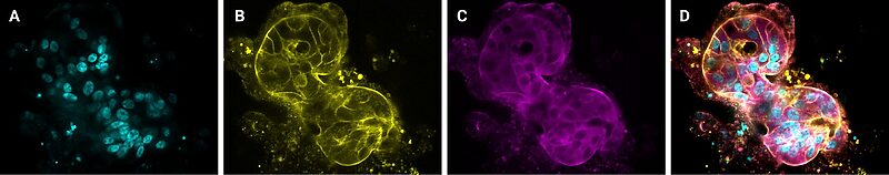
Fig 6. iPSC-derived intestinal organoids cultured and stained in VitroPrime™ 3D Culture and Imaging Plate.
Representative images showing iPSC-derived intestinal organoids cultured in VitroGel® ORGANOID within a 96-well VitroPrime™ 3D Culture and Imaging Plate. iPSC cells were initially cultured in VitroGel® STEM for spheroid and subsequently differentiated into intestinal organoids using CytoGrow™ growth factors and RocketCell™ xeno-free media. Once matured, organoids were fixed and stained without disturbing cultures in the VitroPrime™ 3D Culture and Imaging Plate. Imaging was using a Leica Mica confocal microscope. (A) Blue staining (DAPI) represents cell nuclei (B). Yellow fluorescence: Phalloidin (C), Magenta: Villin protein. (D) Merged image.
Enhance your 3D cell culture with these products:
Hydrogels - Ready-To-Use
ready-to-use, xeno-free (animal origin-free) hydrogel system for organoid culture
Hydrogels - Ready-To-Use
ready-to-use, xeno-free (animal origin-free) hydrogel system for hPSCs 3D static suspension culture and scale-up
New
Cell Culture Medium (Xeno-Free)
An all-in-one xeno-free kit with optimized matrix, medium, and reagents for 3D iPSC expansion.
New
Cell Culture Medium (Xeno-Free)
Advanced xeno-free essential core medium for organoid development that eliminates the need for supplemental additives such as N2 and B27.
New
Cell Culture Medium (Xeno-Free)
Xeno-free medium for 3D expansion of pluripotent stem cells

Downstream Cell Analysis
Fast (15 min), versatile, live/dead cell viability analysis for 3D and 2D cell culture. IN STOCK
| Plate Size | 6-well, 24-well, 96-well |
|---|---|
| Pack Size | 5 Packs/Case |
Related products

Culture Plates
Unique surface treated for superior hydrogel spreading, adherence, and uniform surface.
New
Culture Plates
Premium U-bottom cell culture plate for 3D spheroids.
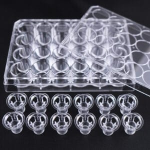
3D Culture Vessels
Transparent, PET, Sterile, 48 inserts (12/PK, 4PK/CS)