Author Archives: Kyra Baricaua

SfN Neuroscience 2025 Recap: A Week of Innovation, Collaboration, and Discovery
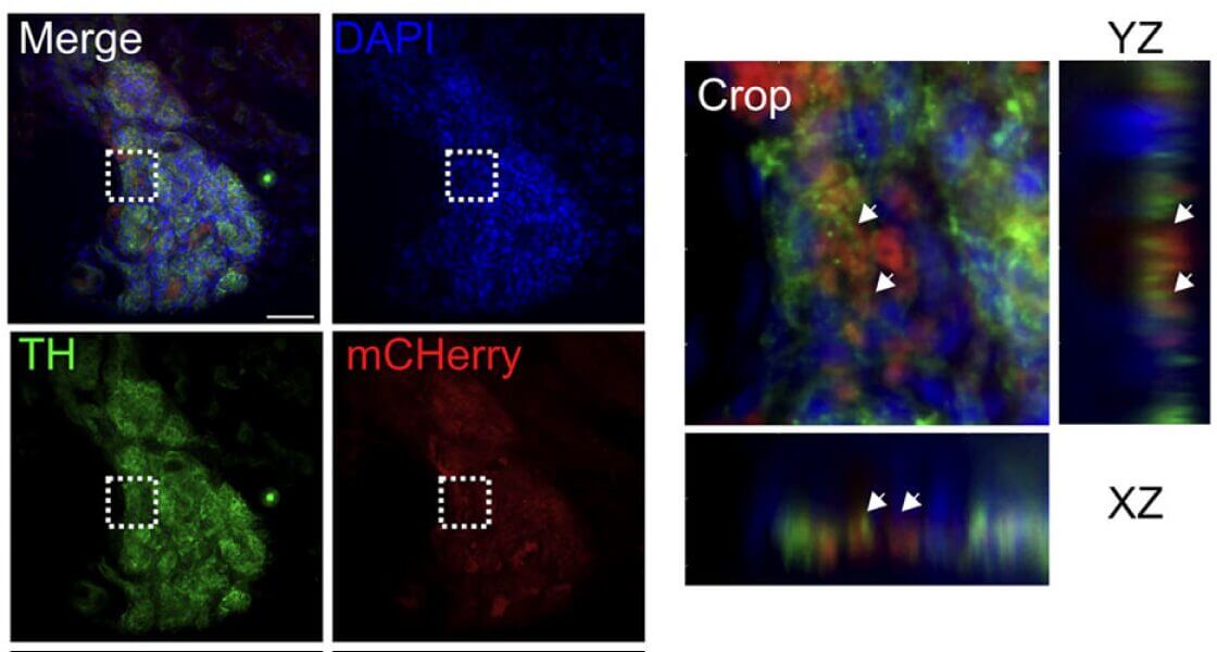
VitroGel® 3D enables precise chemogenetic targeting of carotid body cells to uncover metabolic function. During intense exercise, our bodies have a “ceiling” for oxygen consumption, known as peak oxygen uptake (VO₂ peak), which is a key determinant of athletic endurance. For decades, scientists have known that a tiny organ in the neck, the carotid body, […]
VitroGel® MSC overcomes the limitations of 2D and microcarrier systems by delivering a reproducible, scalable, and functional workflow for MSC biomanufacturing, from expansion to genetic engineering. The production of mesenchymal stem cells (MSCs) at a clinical scale faces persistent challenges. Traditional 2D culture systems and microcarriers are limited by scalability, high costs, and difficulties in […]
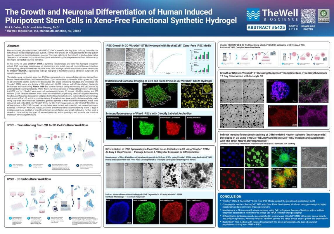
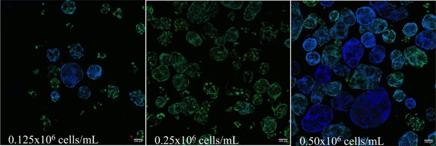
Abstract: Human induced pluripotent stem cells (iPSCs) offer a powerful starting point to study the molecular dynamics of the developing nervous system. Further, they provide an invaluable tool for developing patient-specific models to study the disease process. Therefore, optimizing their growth in a defined, native 3D state is of paramount importance to both guide and […]
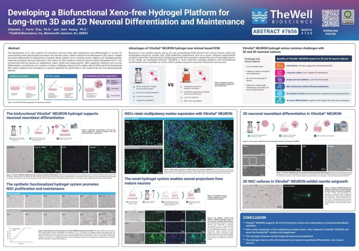
Abstract: The development of in vitro systems for long-term neuronal differentiation and survival is crucial for understanding both physiological processes and various disease states. While traditional two-dimensional (2D) neuron models have been instrumental in neuroscience research, these systems fail to maintain neuron viability over extended periods, impeding prolonged neuronal maturation. One reason for this limitation is that 2D-based models misrepresent […]
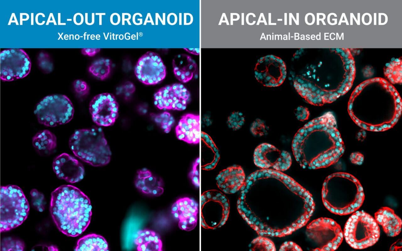
Epithelial polarity, the organized distinction between an apical surface (that faces lumen or the outside) and a basolateral surface (that interfaces with stroma and blood) is a fundamental feature of many tissues, especially the intestinal epithelium. In recent years, three-dimensional (3D) organoid models have become central to nutrition, pharmacology, infection biology, and personalized medicine because […]
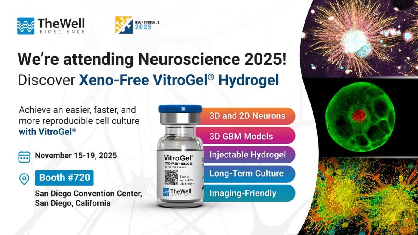
Discover the Future of 3D Cell Culture at SfN – Neuroscience 2025 We’re excited to announce our first-ever participation at Neuroscience 2025! Join us on November 15–19, 2025, at the San Diego Convention Center to explore how VitroGel®, our xeno-free, synthetic hydrogel system, is driving the shift toward animal-free, reproducible, and scalable 3D cell culture […]
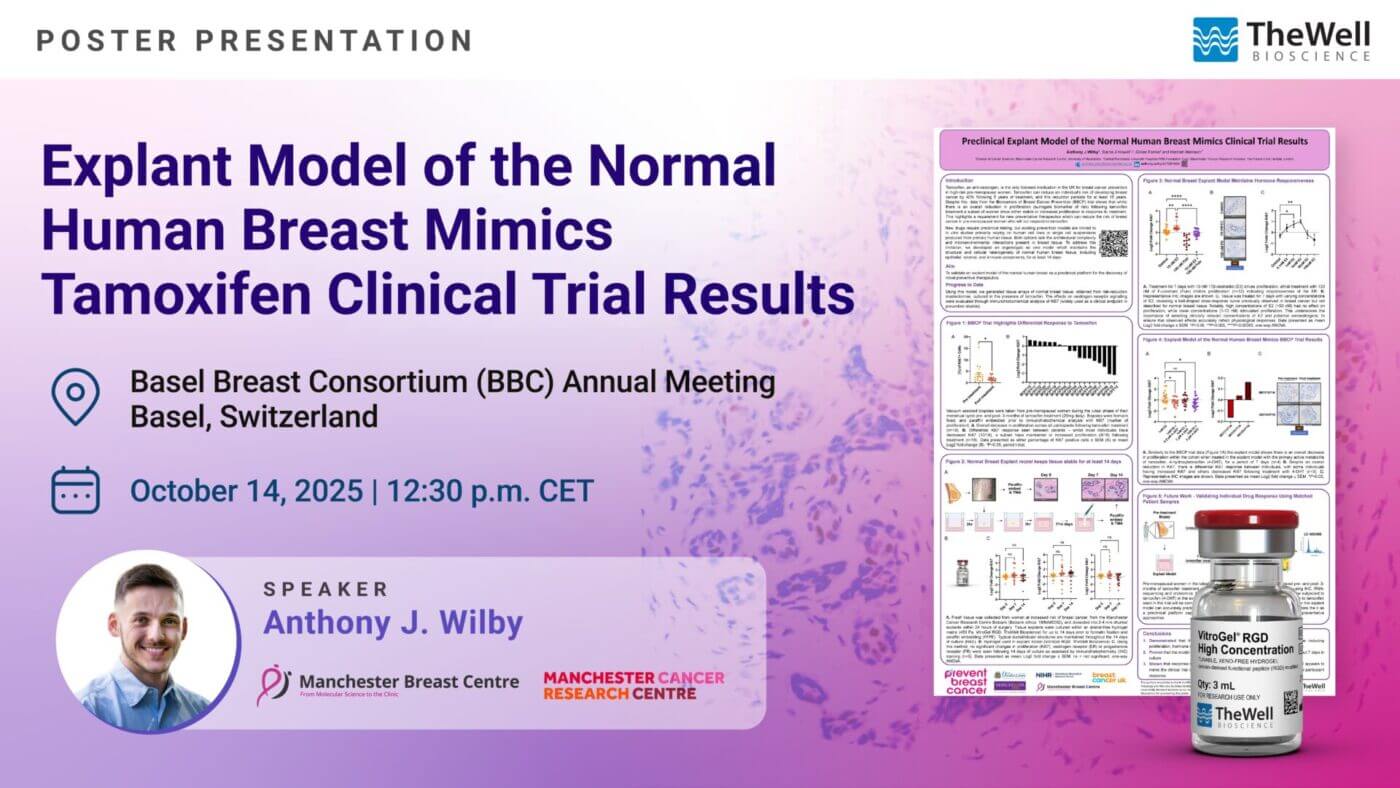
Abstract: Tamoxifen, an anti-oestrogen, is the only licensed medication in the UK for breast cancer prevention in high-risk, pre-menopausal women. Data from the Biomarkers of Breast Cancer Prevention (BBCP) trial show that, while tamoxifen reduces overall proliferation after three months of treatment, a subset of women (5/13) show stable or increased proliferation. As proliferation is […]
Uncovering the power of fibroblast-derived migrasomes in wound healing with the help of VitroGel® 3D’s advanced 3D culture system Effective skin wound healing requires precise cell–cell communication and coordination across tissue types. However, the role of newly discovered migrasomes—vesicular organelles derived from fibroblasts—was not fully understood in the context of tissue repair. To study these […]