Xeno-free 3D Tumoroid Platform
Complete animal-free system for 3D GBM tumoroid generation with batch-to-batch consistency.
NEW
VitroGel® Glioblastoma (GBM) Xeno-Free EMT Kit
Ready-to-use kit for 3D GBM tumoroid formation.
Supports epithelial-to-mesenchymal transition with long-term tumoroid culture.
VitroGel® VitroGel Glioblastoma (GBM) Xeno-Free EMT Kit
Ready-to-use kit for 3D GBM tumoroid formation, supporting epithelial-to-mesenchymal transition and enabling long-term tumoroid culture.

Simple Workflow at Room Temperature
Generate the tumoroid from a simple, single spheroid with an easy operating protocol at room temperature.

Enables EMT Process and Long-Term Culture
Supports the biologically relevant epithelial-to-mesenchymal transition and maintain the well-organized structure for months.

Automation-Friendly and HTS
Compatible with lab-automated liquid handling systems and supports high-throughput screening process.
The VitroGel® Glioblastoma (GBM) Xeno-Free EMT Kit is an excellent tool for generating 3D GBM tumoroids, enabling cells to undergo the epithelial-to-mesenchymal transition for examining pathways involved in cancer metastasis, drug screening, and high-throughput automation.
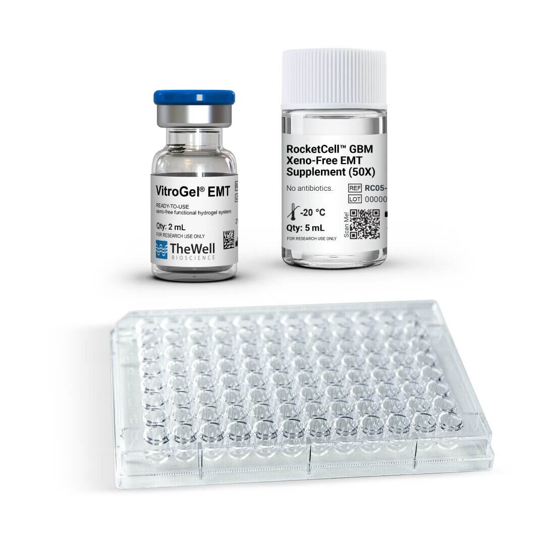
The kit features the ready-to-use and synthetic VitroGel® EMT hydrogel, RocketCell™ GBM Xeno-Free EMT supplement (50X), and VitroPrime™ Ultra-Low Attachment, U-Bottom, 96-Well Plate.
The VitroGel® Glioblastoma (GBM) Xeno-Free EMT Kit is designed to induce tumoroid formation from a simple single spheroid to generate a biologically relevant and well-organized tumoroid model for studying tumor progression, EMT dynamics, and therapeutic response in a preclinical setting.
VitroGel® Glioblastoma (GBM) Xeno-Free EMT Kit (VHM08-K)
Contents:
- 1 x 2 mL, VitroGel® EMT hydrogel
- 1 x 5 mL, RocketCell™ GBM Xeno-Free EMT supplement (50X)
- 1 x VitroPrime™ Ultra-Low Attachment Plate, U-Bottom, 96-Well
VitroGel® Glioblastoma (GBM) Xeno-Free EMT High-Screening Kit
(VHM08-HK) Contents:
- 5 x 2 mL, VitroGel® EMT hydrogel
- 5 x 5 mL, RocketCell™ GBM Xeno-Free EMT supplement (50X)
- 5 x VitroPrime™ Ultra-Low Attachment Plate, U-Bottom, 96-Well
Specifications
| Use | 3D Glioblastoma tumoroid formation for drug screening purposes |
| Foormulation | Xeno-free, biofunctional hydrogel and supplement system |
| Injectability | Injectable hydrogel for in vivo studies and lab automation |
| Operation | Ready-to-use at room temperature |
| Cell Harvesting | VitroGel® Organoid Recovery Solution 5-15 min cell recovery |
| Shipping and Storage | Hydrogel: Ships at ambient temperature. Store at 2-8°C. Supplement: Dry Ice shipping. Store at -20°C. |
Challenges We Solve

How VitroGel® GBM Xeno-Free EMT Kit


Common Challenges in 3D Tumoroid Generation
Lack of a robust 3D EMT tumoroid model tounderstand cancer metastasis
Hard to support a long-term
tumoroid cultures
Lack of a method to establish comprehensive tumoroid models from well characterized cell lines
Difficult to adapt lab-automation and high-throughput screening for big-sized 3D tumoroids
How VitroGel® GBM Xeno-Free EMT Kit
Solves The Issues
Induce a controllable EMT process for tumoroid formation
The unique combination of VitroGel® EMT hydrogel and RocketCell™ EMT supplement induces the epithelial-to-mesenchymal transition for glioblastoma cells.
Long-term Culture
Over 6 weeks of tumoroid maintenance for prolonged studies.
Simple, Fast, and Reliable
Establish tumoroid formation from well-established cell lines in less than a week with highly reproducible results.
Automation-friendly
Room temperature operation makes an easy transition to high-throughput platforms.
Protocols and Resources
Product Documentation
![]() VitroGel® Glioblastoma (GBM) Xeno-Free EMT Kit – Sale Sheet
VitroGel® Glioblastoma (GBM) Xeno-Free EMT Kit – Sale Sheet
![]() Product Data Sheet
Product Data Sheet
Data and References
CASE 1
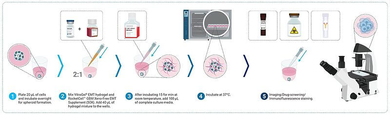
3D Tumoroid formation with the VitroGel® Glioblastoma Xeno-Free EMT Kit
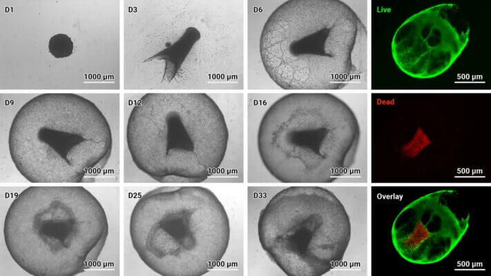
Figure 1: VitroGel® Glioblastoma Xeno-Free EMT kit supports tumoroid formation.
U87 MG cells were resuspended in basal medium supplemented with 1X RocketCell™ GBM Xeno-Free EMT supplement and seeded into the VitroPrime™ Ultra-Low Attachment Plate, U-Bottom, 96-Well, to promote overnight spheroid formation. The spheroids were supplemented with VitroGel® EMT and RocketCell™ GBM Xeno-Free EMT supplement to induce tumoroid generation. Light microscopy was performed multiple days to evaluate tumoroid growth over time (left). Cyto3D® Live-Dead Assay staining was performed on GBM tumoroids. Live cells within the tumoroid are shown as green and dead cells in red (right).
CASE 2
Evaluation Viability of GBM Tumoroids
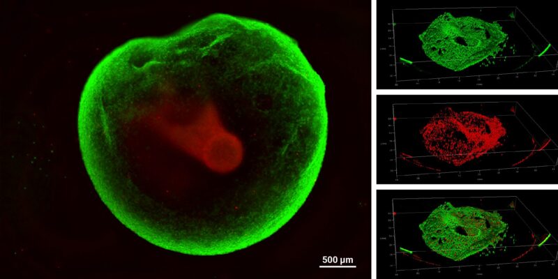
Figure 2: VitroGel® Glioblastoma Xeno-Free EMT kit hydrogel sustains tumoroid growth and viability.
GBM tumoroids were subjected to cell viability staining using Cyto3D® Live-Dead Assay Kit after six weeks in culture. Live cells within the tumoroid are shown in green, and dead cells are shown in red.
CASE 3
Tumoroids grown with the VitroGel® Glioblastoma Xeno-Free EMT Kit
as a Platform for Drug Screening
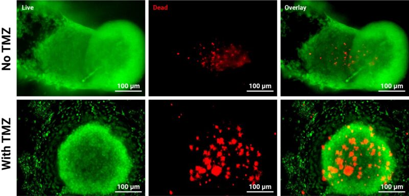
Figure 3: GBM tumoroids are susceptible to chemotherapy.
Tumoroids were grown for 3 days in VitroGel® EMT hydrogel with RocketCell™ supplement system. The tumoroids were treated with Temozolomide (TMZ; 1mM) for 24 hours and subjected to cell viability studies. The Cyto3D® Live-Dead Assay Kit was used to label live cells (green) and dead cells (red) in the tumoroid, both with and without drug treatment. The pictures were obtained at a 20X magnification.
CASE 4
Tumoroid Characterization: Evaluation of EMT Markers
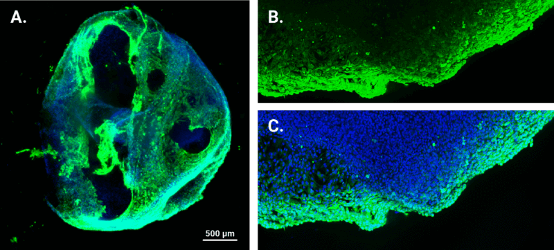
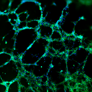
Figure 4: Assessing Vimentin expression, an EMT marker, in GBM tumoroids.
Immunofluorescence staining was employed on three-week-old tumoroids to examine Vimentin expression. The nuclei were stained with DAPI (blue), and vimentin was stained green. A. Representative image of tumoroid topology and size (4X magnification). B-C. Enlarged images of vimentin-positive regions obtained with a confocal microscope at 10X magnification.
| Kit | 1 Kit, 5 Kits |
|---|
Related products
Assay Kits
Tunable hydrogel system for 2D gel coating and 3D culture of angiogenesis tube formation, invasion, and animal injection.
Xeno-free hydrogel system for consistent study of cell invasion. Kit includes choice of tunable functionalized hydrogel, 10mL VitroGel® Dilution Solution TYPE 2, and VitroPrime™ Cell Culture Inserts, 8 µm.
Assay Kits
ready-to-use, hydrogel system for 2D gel coating and 3D culture of angiogenesis tube formation, invasion, and animal injection.
Assay Kits
A simple and easy replacement for animal-based ECM for consistent cell invasion studies. This kit includes the VitroGel® Hydrogel Matrix with VitroPrime™ Cell Culture Inserts, 8 µm, PET