Xeno-free and Reproducible
Complete animal-free system for spheroid formation, tumoroid culture with batch-to-batch consistency.
NEW

VitroGel® EMT
Xeno-free hydrogel that supports spheroid formation, tumoroid culture, and uniquely enables EMT biology
Full xeno-free EMT kit available. Click here
VitroGel® EMT
Ready-to-use hydrogel for spheroid formation, tumoroid culture, and uniquely enables EMT biology
Simple Workflow at Room Temperature
Generate the tumoroid from a simple, single spheroid with an easy operating protocol at room temperature.
Enables EMT Process and Long-Term Culture
Supports the biologically relevant epithelial-to-mesenchymal transition and maintain the well-organized structure for months.
Automation-Friendly and HTS
Compatible with lab-automated liquid handling systems and supports high-throughput screening process.
VitroGel® EMT is a fully defined, xeno-free hydrogel engineered as a protocol-compatible alternative to low-concentration Matrigel® (5%) for spheroid, tumoroid, and organoid formation.
Designed to integrate seamlessly into existing workflows without requiring protocol modifications, VitroGel® EMT enables researchers to transition away from animal-derived matrices while maintaining robust 3D culture performance.
Beyond supporting reproducible spheroid generation, VitroGel® EMT provides a controlled microenvironment for epithelial–mesenchymal transition (EMT) studies, enabling researchers to investigate tumor progression, invasion, metastasis, and therapeutic response with greater experimental consistency.
As part of a scalable 3D culture platform, VitroGel® EMT supports applications ranging from model development and disease biology to high-throughput drug screening and translational research.
Specifications
| Formulation | Xeno-free, biofunctional hydrogel |
| Use | spheroid formation, tumoroid culture, and uniquely enables EMT biology |
| Operation | Ready-to-use at room temperature |
| Injection | Injectable hydrogel for in vivo studies and lab automation |
| pH | Neutral |
| Storage | Store at 2-8°C. Ships at ambient temperature |
| Sizes | 10 mL and 2 mL |
Challenges We Solve


Challenges when using animal-based ECM

Lot-to-lot vairability undermines reproducibility

ECM protein derived from a mouse tumor; ~2000 undefined compounds, including various growth factors. Ethical- concerns

Cold-chain dependency, must be kept on ice at all times
No EMT biology, passive matrix with no mechanistic added value.

Cost and supply chain vulnerabilities.
What VitroGel® EMT delivers
Batch-to-batch consistency.
Synthetic and defined formulation.
100% Xeno-free; no animal-derived components
Room temperature operation.
No cold-chain hassle.
EMT-active.
Uniquely supports EMT biology.

No protocol changes.
Same as 5% workflow, drop-in replacement.
Protocols and Resources
![]() VitroGel® EMT hydrogel – Protocol
VitroGel® EMT hydrogel – Protocol
Product Documentation
![]() VitroGel® EMT hydrogel – Sale Sheet
VitroGel® EMT hydrogel – Sale Sheet
![]() Product Data Sheet
Product Data Sheet
![]() Material Safety Data Sheet (MSDS)
Material Safety Data Sheet (MSDS)
Data and References

CASE 1
3D Tumoroid formation: VitroGel® EMT vs. Matrigel

Figure 1: GBM tumoroid development in VitroGel® EMT hydrogel system. U87-MG GBM cells (1 x 10⁶ cells/mL) were resuspended in basal medium with supplements. 20 µL of cell suspension was added to the VitroPrime™ Ultra-Low Attachment, U-Bottom, 96-Well Plate and incubated overnight at 37°C. VitroGel® EMT hydrogel was mixed with RocketCell™ GBM Xeno-Free EMT supplement in a 1:1 ratio, and 40 µL of the mixture was added to the cultures. The hydrogel was incubated at room temperature for 15 minutes. A 100 µL of basal medium with supplements was added on top of the hydrogels. In parallel, Matrigel was diluted 1:10 with basal medium containing FBS. The cultures were incubated at 37°C. Tumoroid growth was monitored for 25 days and evaluated using a Zeiss microscope.
CASE 2
Evaluation Viability of GBM Tumoroids
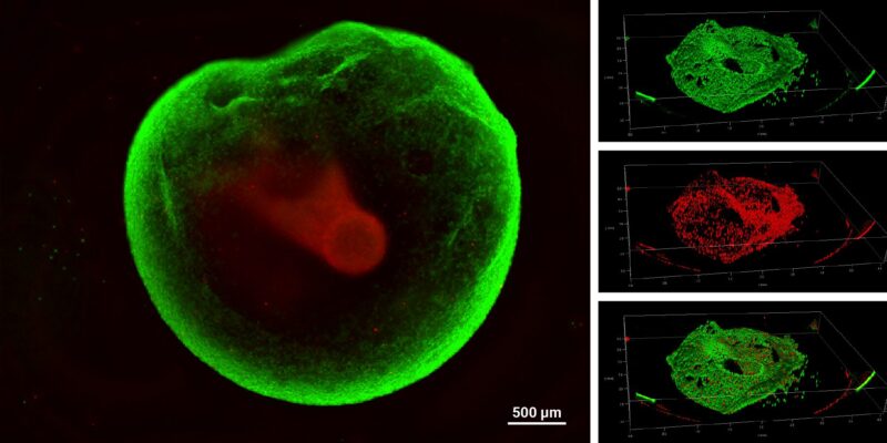
Figure 2: VitroGel® Glioblastoma Xeno-Free EMT kit hydrogel sustains tumoroid growth and viability.
GBM tumoroids were subjected to cell viability staining using Cyto3D® Live-Dead Assay Kit after six weeks in culture. Live cells within the tumoroid are shown in green, and dead cells are shown in red.
CASE 3
Tumoroids grown with the VitroGel® Glioblastoma Xeno-Free EMT Kit
as a Platform for Drug Screening
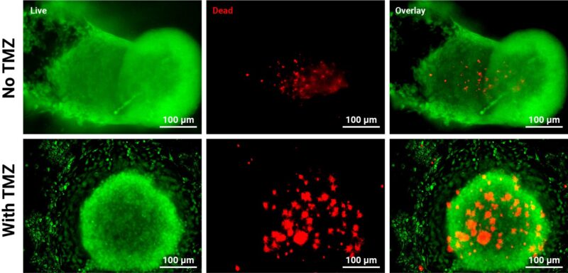
Figure 3: GBM tumoroids are susceptible to chemotherapy.
Tumoroids were grown for 3 days in VitroGel® EMT hydrogel with RocketCell™ supplement system. The tumoroids were treated with Temozolomide (TMZ; 1mM) for 24 hours and subjected to cell viability studies. The Cyto3D® Live-Dead Assay Kit was used to label live cells (green) and dead cells (red) in the tumoroid, both with and without drug treatment. The pictures were obtained at a 20X magnification.
CASE 4
Tumoroid Characterization: Evaluation of EMT Markers
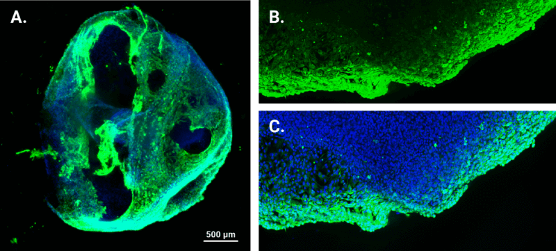
Figure 4: Assessing Vimentin expression, an EMT marker, in GBM tumoroids.
Immunofluorescence staining was employed on three-week-old tumoroids to examine Vimentin expression. The nuclei were stained with DAPI (blue), and vimentin was stained green. A. Representative image of tumoroid topology and size (4X magnification). B-C. Enlarged images of vimentin-positive regions obtained with a confocal microscope at 10X magnification.
Resources

| Size | 2 mL, 10 mL |
|---|
Related products
Hydrogels - Ready-To-Use
ready-to-use, xeno-free (animal origin-free) hydrogel system for hPSCs 3D static suspension culture and scale-up

Hydrogels - Ready-To-Use
ready-to-use, xeno-free (animal origin-free) hydrogel system for organoid culture
NEW
Hydrogels - Ready-To-Use
Ready-to-use, xeno-free hydrogel system for neural stem cell and neuron cultures.
Hydrogels - Ready-To-Use
ready-to-use, xeno-free (animal origin-free) hydrogel system for mesenchymal stem cell 3D culture and scale-up
Hydrogels - Ready-To-Use
ready-to-use, xeno-free (animal origin-free) hydrogel system for 3D culture of HEK293 cells
Hydrogels - Ready-To-Use
ready-to-use, xeno-free (animal origin-free) biofunctional hydrogel system