
N2B27-Premix Animal Origin-Free Supplement
NEW
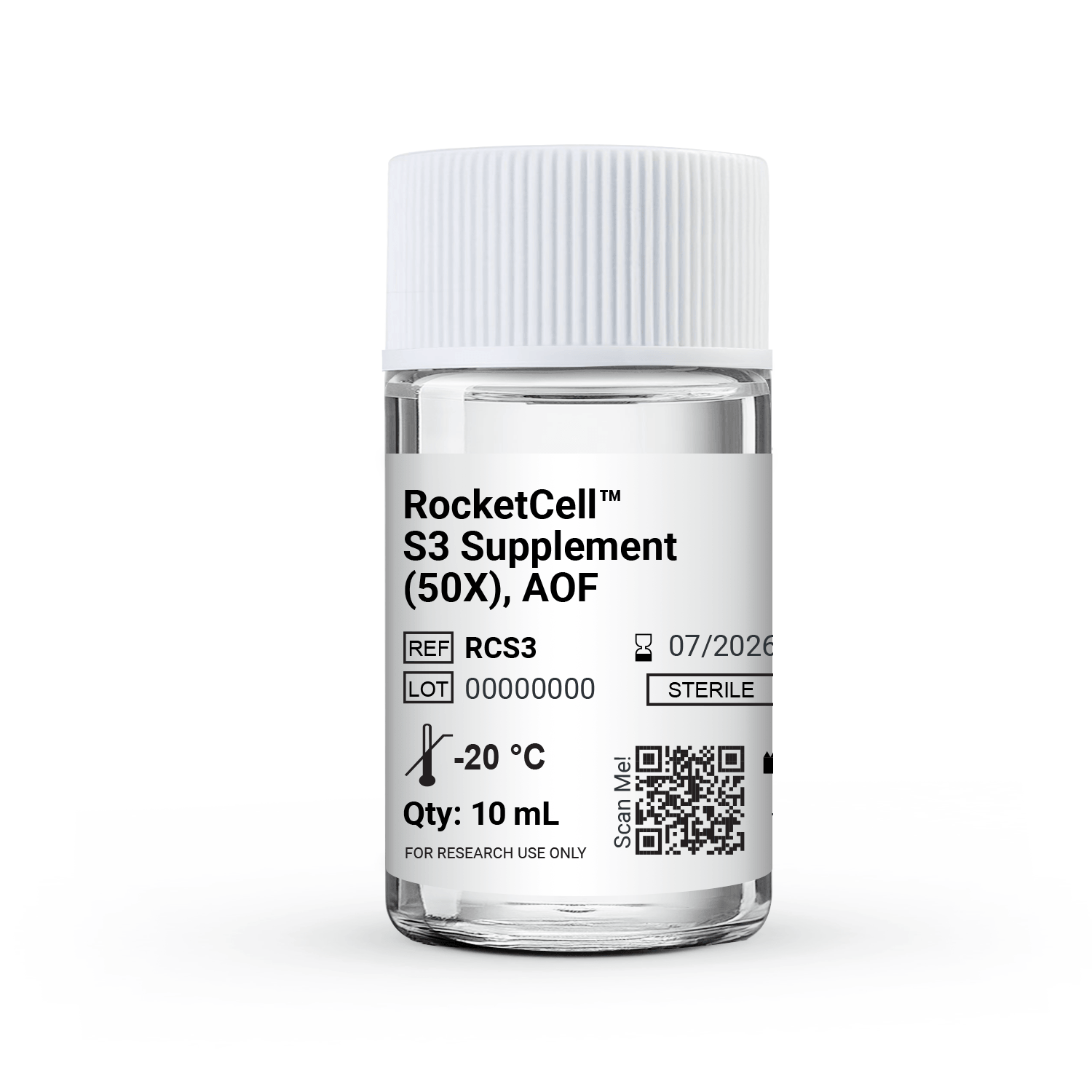
RocketCell™ S3 Supplement (50X), AOF
N2B27-Premix Animal Origin-Free Supplement
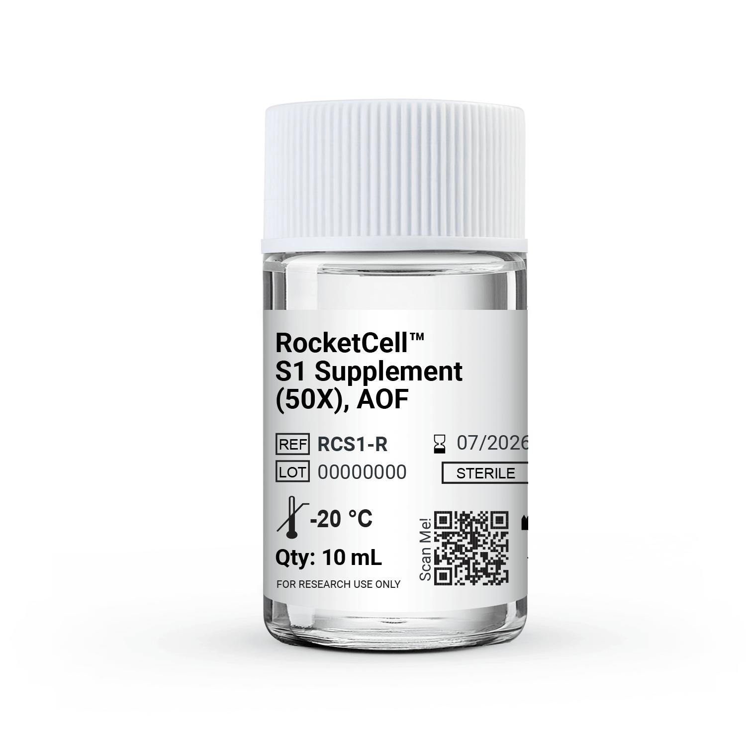
RocketCell™ S3 Supplement (50X), AOF

Synergistic Dual-Supplement Performance
Combining the complementary strengths of both formulations delivers superior cell viability and performance compared to either supplement used alone.

3D and Co-culture Compatible
Formulated for stability and performance within VitroGel® hydrogel-based 3D culture systems, supporting complex co-culture and organoid model development.
Also works with animal-based ECMs.

Consistent Lot-to-Lot Quality
Selectively supports post mitotic neurons in mixed primary cultures while suppressing overgrowth of non-neuronal cell populations.

Ready-to-use Convenience
Eliminates the manual mixing step, reducing pipetting variability and preparation time in high-throughput or time-sensitive workflows.

Broad Application Range
Supports neural stem cell expansion, iPSC-derived neuron maturation, cancer stem cell maintenance, and organoid and tumoroid culture across multiple tissue types including intestinal, colonic, pancreatic, lung, retinal, and liver models.
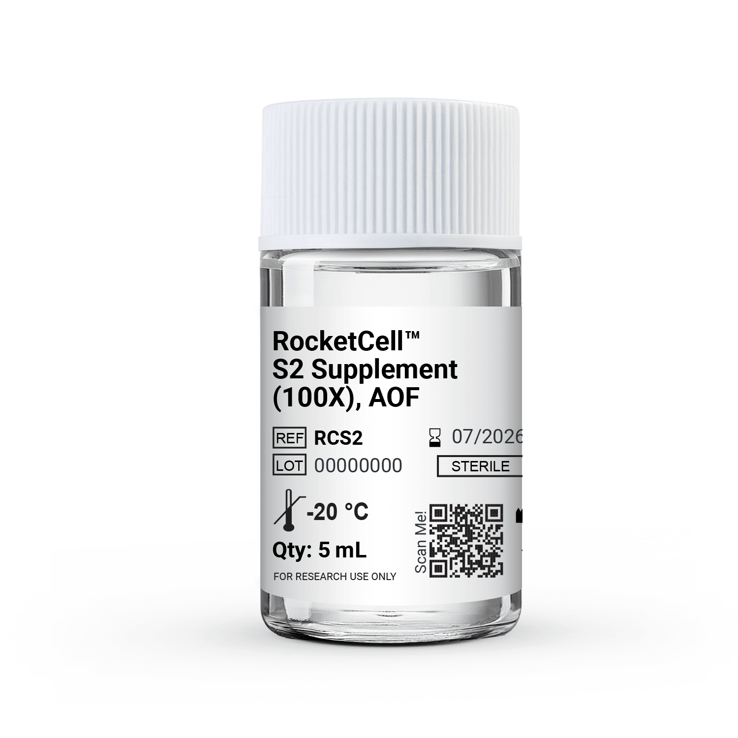
RocketCell™ S3 Supplement, AOF (N2B27) is animal origin-free, ready-to-use, 1:1 premix of the RocketCell™ S2 Supplement (N2-like) and S1 Supplement (B27-like) formulations — a single-component solution for culture paradigms that require both supplements simultaneously.
The combination of N2 and B27 is a cornerstone of modern serum-free culture, appearing in media formulations across an exceptionally broad range of applications including neural, epithelial, and cancer models, as well as organoid and tumoroid systems across multiple tissue types. RocketCell™ S3 Supplement, AOF delivers this established dual-supplement framework in a fully animal origin-free and defined format.
Challenges We Solve


Common Challenges

Lack of a reliable animal origin-free formulation to support GMP process.

Multi-supplement media preparation increases preparation time and handling steps.

Variability in media preparation when multiple supplements are mixed manually.
Limited availability of ready-to-use N2/B27-like supplements for diverse organoid models.
How RocketCell™ S3 Supplement, AOF Solves The Issues
Animal origin-free formulation for GMP transition
N2/B27-like AOF formulation that simply the transition to
scalable and controlled GMP workflows.
Simplified Media Preparation
Combines N2- and B27-like components in one bottle, reducing media prep time, pipetting steps, and contamination risk.
Consistent Supplementation
Delivers a standardized N2B27-like formulation across
experiments and workflows.
Robust Formulation for Organoid Applications
Supports neural stem cell expansion, iPSC-derived neuron maturation, cancer stem cell maintenance, and organoid/tumoroid culture across multiple tissue types.
Protocols and Resources
 Protocol

Data and References
iPSC-derived Intestinal Organoids Cultured Using RocketCell™ S3 (N2B27) Supplement, AOF
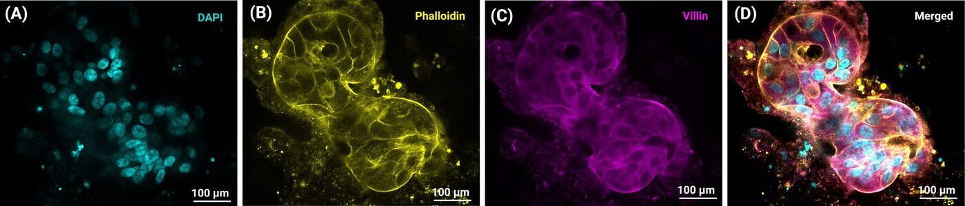
Use of VitroGel® hydrogels & RocketCell™ S3 Supplement, AOF to generate iPSC-derived intestinal organoids with apical-out polarity.
Representative image showing iPSC derived intestinal organoids cultured in VitroGel® ORGANOID. iPSC cells were cultured in VitroGel® STEM to generation of spheroids cells. These spheroids were then differentiated to intestinal organoids using organoid culture media formulated using RocketCell™S3 Supplement, AOF and CytoGrow™ growth factors. (A) Blue staining (DAPI) represents cell nuclei. (B) Yellow fluorescence: Phalloidin, (C) Magenta: Villin protein, & (D) Merged image.
Generation of iPSC-derived Liver Organoid in RocketCell™ S3 (N2B27) Supplement, AOF and Synthetic VitroGel® ORGANOID Hydrogel
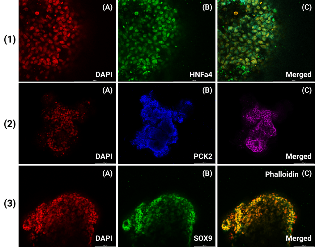
Generation and characterization of iPSC-derived liver organoids using RocketCell™ S3 (N2B27) Supplement and synthetic, chemically defined VitroGel® ORGANOID Hydrogel.
Representative bright-field images demonstrate the formation and progressive development of iPSC-derived liver organoids in 3D culture. Immunofluorescence analysis confirms liver-specific characteristics, including HNF4α expression (1), PCK2 expression and F-actin/Phalloidin staining (2), and SOX9 expression (3). DAPI staining indicates cell nuclei, with merged images showing the corresponding marker expression within the organoid structures.
Mouse Intestinal Organoid Development using
RocketCell™ S3 (N2B27) Supplement, AOF in Animal-based ECM
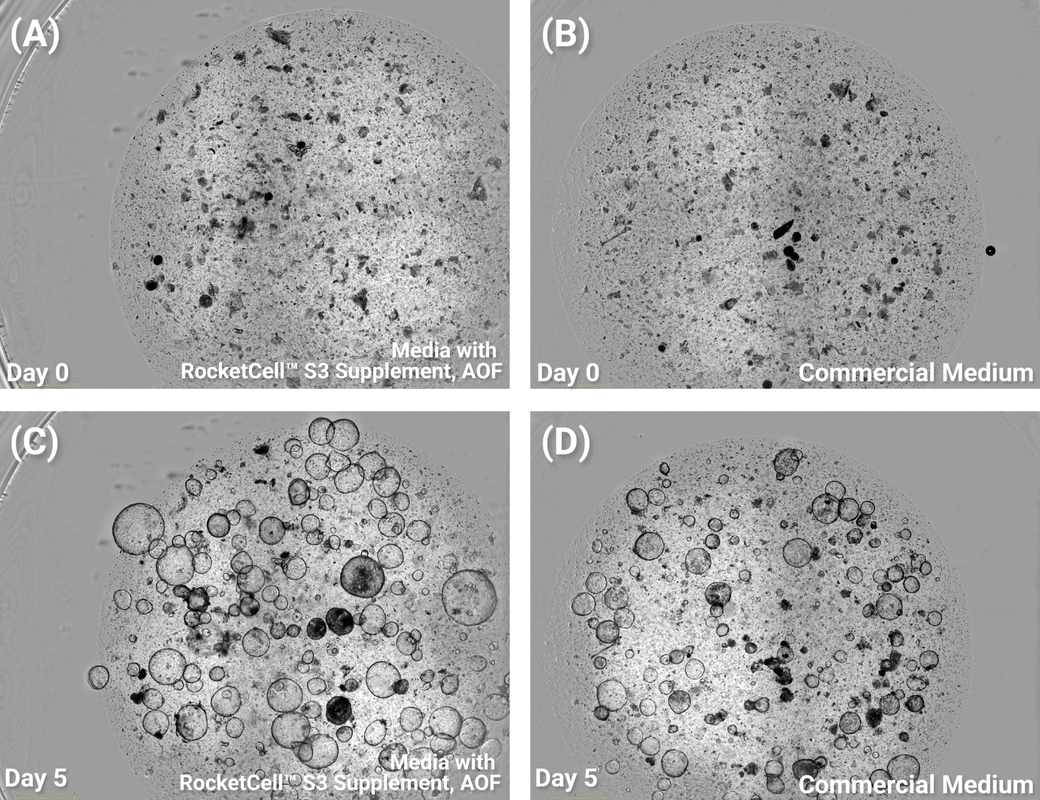
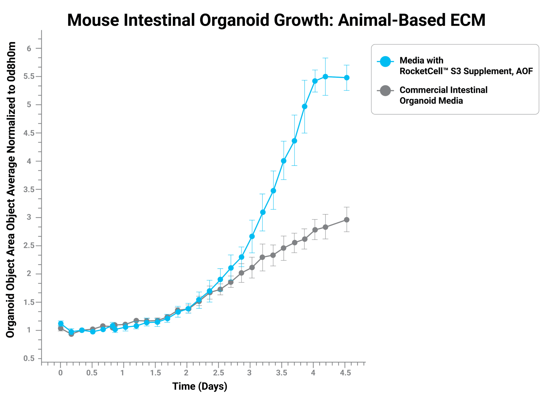
Mouse intestinal organoid growth in animal-based ECM using RocketCell™ S3 Supplement, AOF.
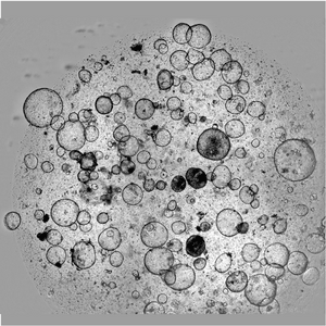
Representative bright-field images show organoid development from Day 0 to Day 5 in RocketCell™-supplemented medium compared with commercial intestinal organoid medium. Quantitative analysis demonstrates enhanced organoid growth with RocketCell™ medium, with greater expansion observed from approximately Day 2 onward.
Differentiated Neurospheres (Brain Organoids) developed in VitroGel® NEURON, RocketCell™ NSC Growth Medium, AOF and RocketCell™ S3 (N2B27) Supplement, AOF
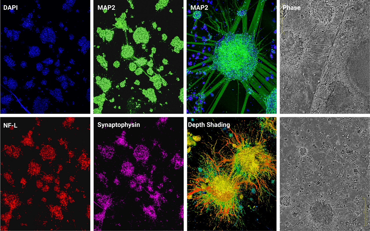
Neurosphere differentiation and culture in VitroGel® NEURON, RocketCell™ NSC Growth Medium, AOF, and RocketCell™ S3 Supplement , AOF
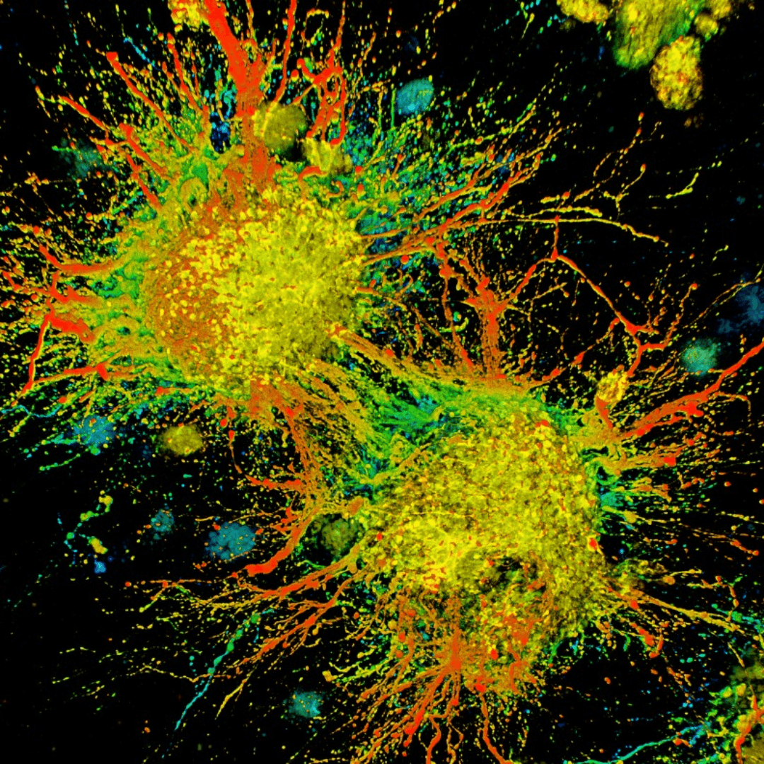
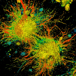
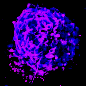
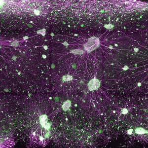
Immunofluorescence staining of differentiated Neuron-Spheres (Brain Organoids) developed in 3D using VitroGel® NEURON and RocketCell™ NSC Growth Medium, AOF in combination with RocketCell™ S3 Supplement, AOF (N2B27 Alternative). The images were obtained with Confocal Microscopy – Maximum Projection and Incucyte S3 Standard 20x Tracking.
Neurospheres (Brain Organoids) Differentiation in VitroGel® NEURON, RocketCell™ NSC Growth Medium, AOF and RocketCell™ S3 (N2B27) Supplement, AOF
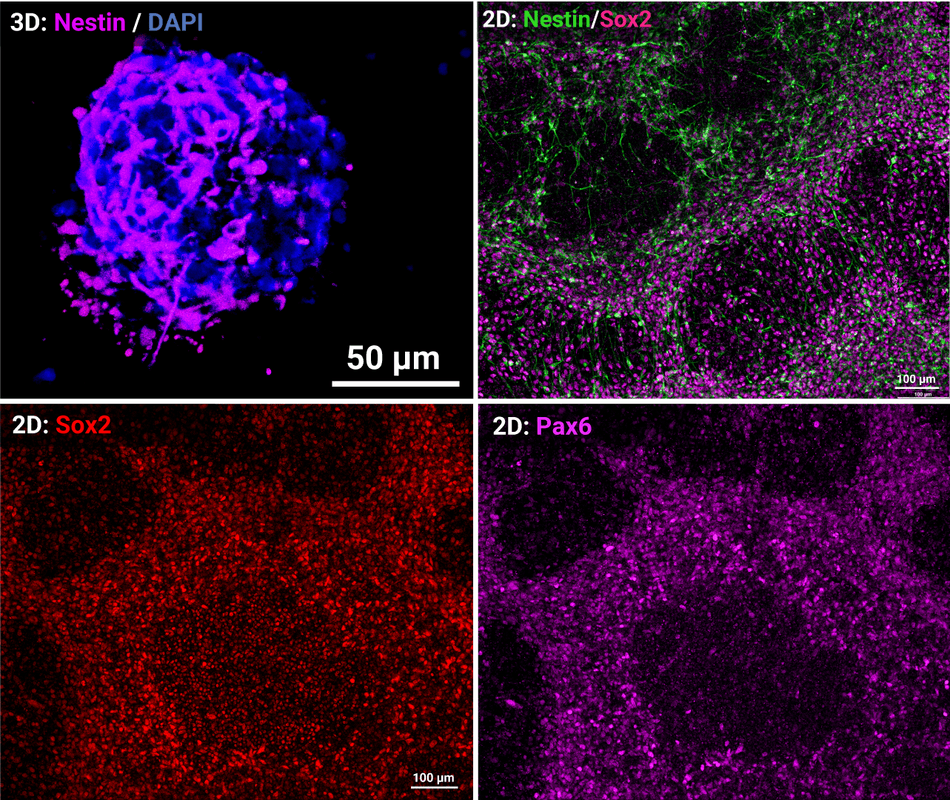
Growth of NSCs in VitroGel® STEM using RocketCell™ NSC Growth Medium, AOF
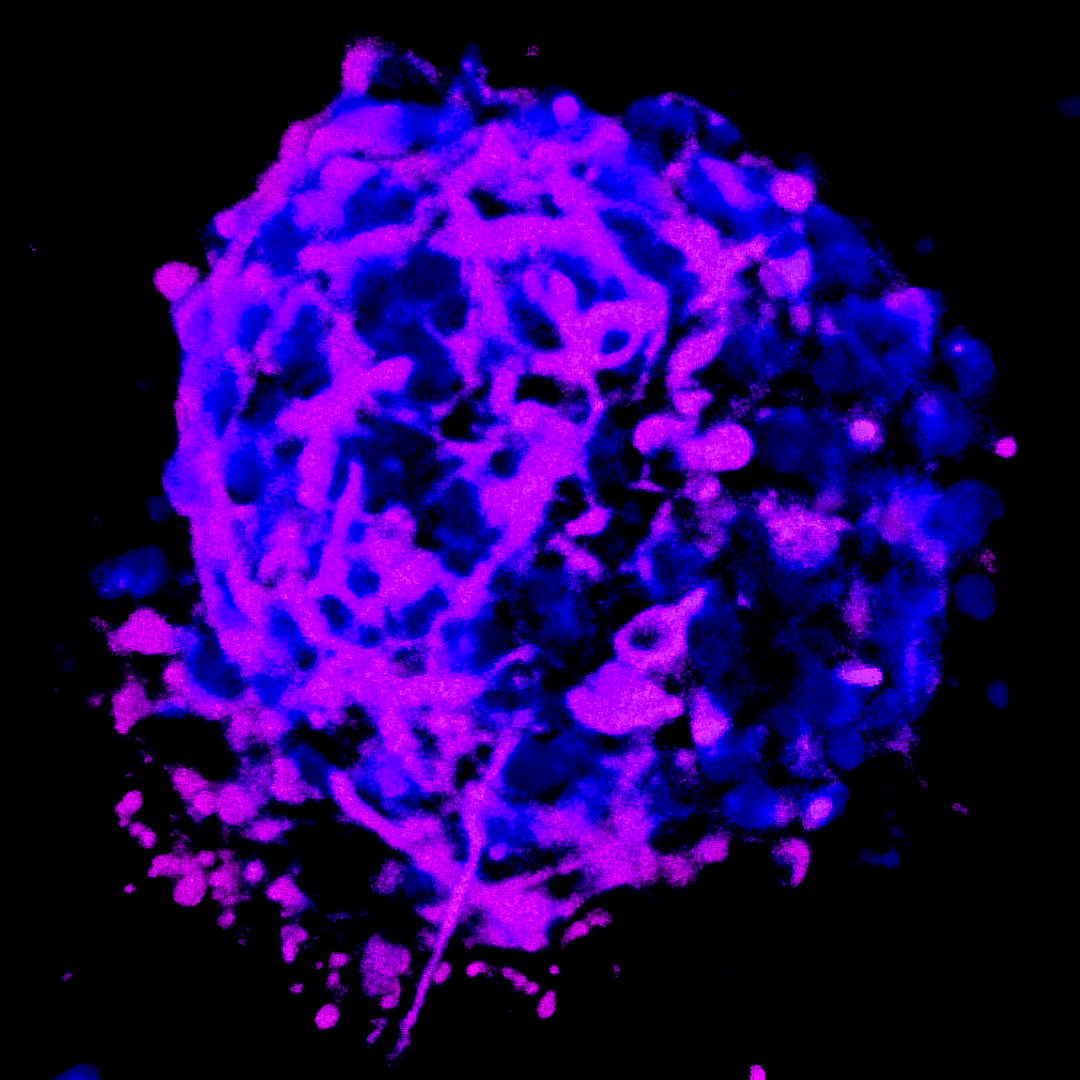
iPSC-derived NSCs were cultured in VitroGel® STEM and the RocketCell™ NSC Growth Medium, AOF supplemented with the RocketCell™ S3 Supplement, AOF (N2B27 Alternative) for 14 days. Immunofluorescence staining depicts expression of NSC-associated markers.
| Size | 10 mL |
|---|
Related products
New
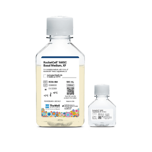

Cell Culture Media (Xeno-Free)
High-performance, xeno-free kit for superior expansion of human mesenchymal stem cells (hMSC)
NEW
Cell Viability Enhancer
Maximize cell recovery and growth during passaging, thawing, and cryopreservation.
New
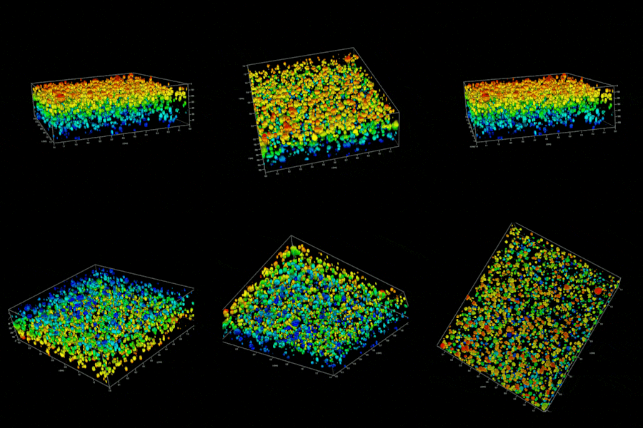
Cell Culture Media (Animal Origin-Free)
An all-in-one, animal origin-free kit with optimized matrix, medium, and reagents for 3D iPSC expansion.
New
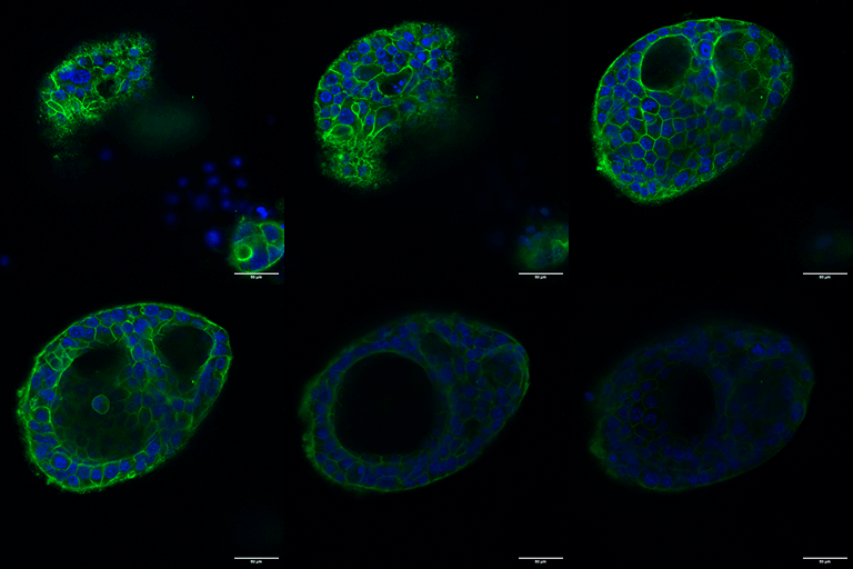
Cell Culture Media (Animal Origin-Free)
All-in-one, animal origin-free kit for growth, expansion, and passaging of apical-out intestinal organoids.
NEW
Culture Media and Supplements
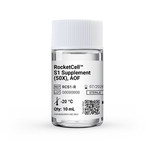
B27-like, Animal Origin-Free Supplement
New
Cell Culture Media (Animal Origin-Free)
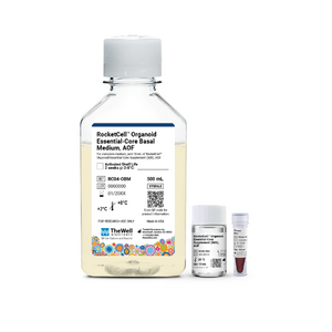
One Core Medium. Endless Organoid Possibilities. An animal origin-free medium that supports multiple organoid types - just add growth factors.