
Xeno-Free Formulation
Supports the development and maintenance of 3D spheroid and tumoroid cultures without FBS or other animal-derived components, providing a more defined alternative to conventional serum-based workflows.
NEW
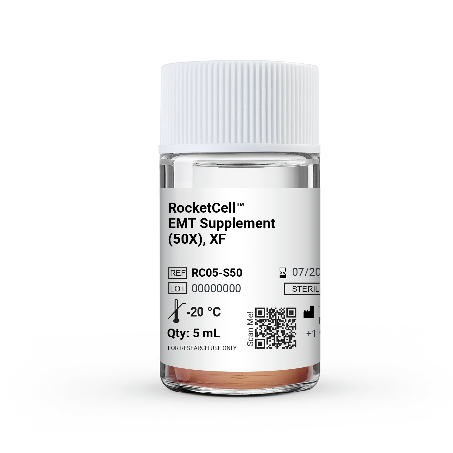
RocketCell™ EMT Supplement (50X), XF
Xeno-Free supplement designed to induce EMT Tumoroid formation.
Hydrogel can be purchased separately. Click here
Full EMT kit available.Click here
$149.00
RocketCell™ EMT Supplement (50X), XF
Xeno-free supplement for 3D spheroid and tumoroid culture

Long-Term 3D Culture
Prolonged spheroid and tumoroid cultures for extended studies of tumor biology and therapeutic response.

Supports Spheroid-to-tumoroid Development
Facilitates continued growth and development of established
spheroids into larger, more complex 3D tumoroids when used
with VitroGel® EMT hydrogel.

Versatile Cancer Research Applications
Supports 3D models for investigating tumor growth and progression, EMT- associated changes, therapeutic response, and drug screening.
RocketCell™ EMT Supplement (50X), XF is a xeno-free supplement designed to support the development and maintenance of 3D spheroid and tumoroid models for cancer research.
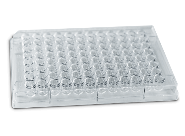
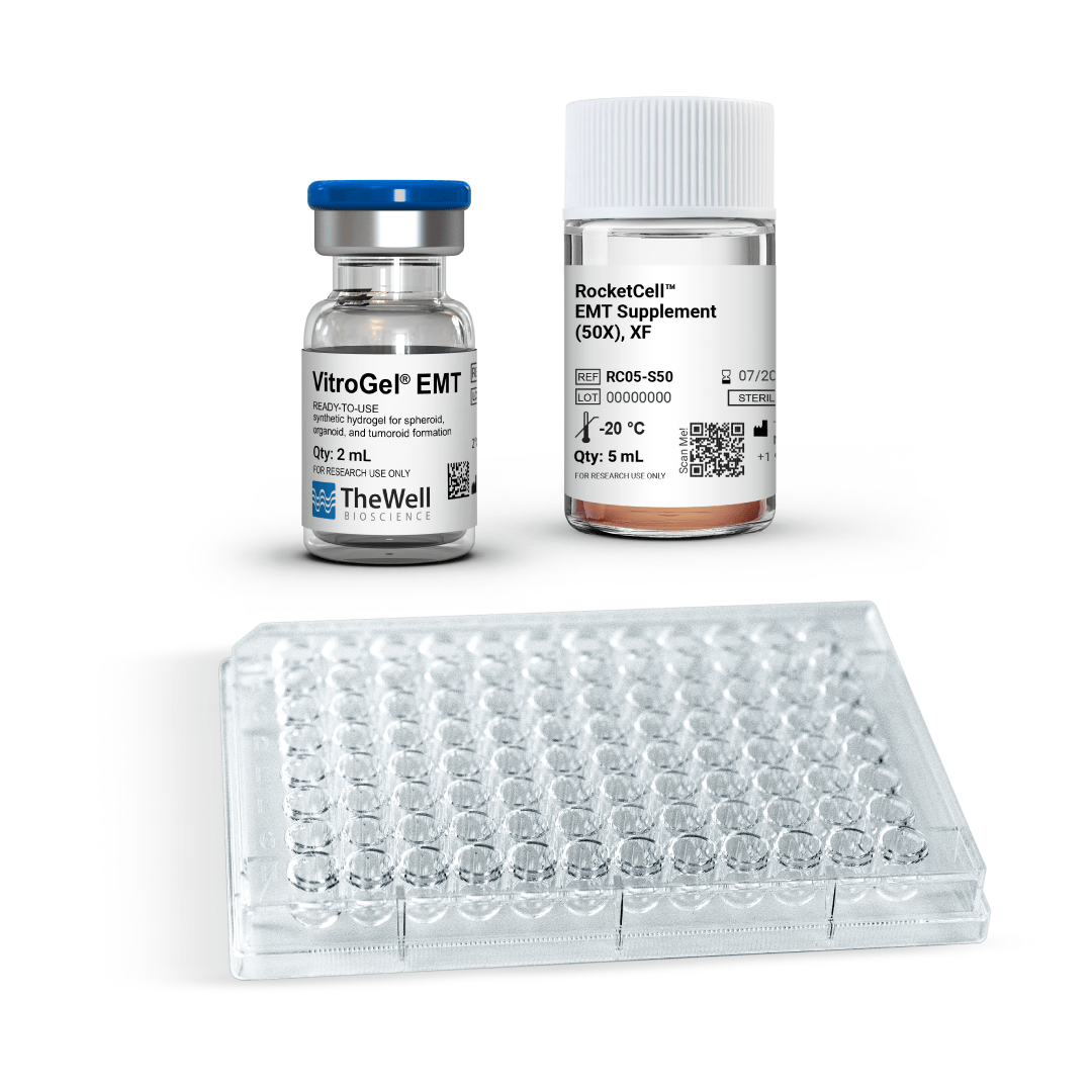
When combined with VitroGel® EMT hydrogel and the VitroPrime™ Ultra-Low Attachment, U-bottom, 96-well plate, the RocketCell™ EMT
Supplement supports continued growth and development of established spheroids into larger, more complex tumoroids.
The resulting 3D models provide a defined platform for studying tumor growth and progression, EMT-associated changes, therapeutic response, and cancer drug screening.
Challenges We Solve
Common


How RocketCell™ EMT Supplement (50X), XF
Common
Challenges

Generating complex 3D tumor models

Sustaining viability in larger 3D models

Establishing defined, xeno-free cultures

Difficult to scale 3D models for high-throughput screening
How RocketCell™ EMT Supplement (50X), XF
Solves The Issues
Supports 3D Tumor Model Development
RocketCell™ EMT Supplement supports the growth and development of spheroid and tumoroid cultures, providing a serum-free environment for establishing complex 3D tumor models.
Long-term 3D Culture Viability
Enables long-term growth and maintains viability of advanced spheroid and tumoroid models.
Enables a Xeno-Free Workflow
The xeno-free formulation supports spheroid and tumoroid culture without FBS or other animal-derived components, providing a defined alternative to conventional culture workflows.

Facilitates High-Throughput Screening
Compatible with high-throughput 3D culture workflows for drug screening and treatment-response studies.
Protocols and Resources


Data and References
tumoroid applications
CASE 1
3D Tumoroid formation with the RocketCell™ EMT Supplement (50X), XF
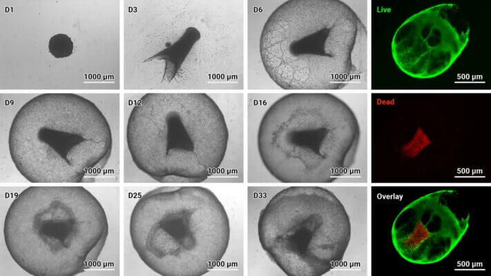
Figure 1: VitroGel® Glioblastoma EMT Kit, XF supports tumoroid formation.
U87 MG cells were resuspended in basal medium supplemented with 1X RocketCell™ EMT Supplement, XF, and seeded into the VitroPrime™ Ultra-Low Attachment Plate, U-Bottom, 96-Well, to promote overnight spheroid formation.
The spheroids were supplemented with VitroGel® EMT and RocketCell™ EMT Supplement, XF to induce tumoroid generation. Light microscopy was performed over multiple days to evaluate tumoroid growth over time (left). Cyto3D® Live-Dead Assay staining was performed on GBM tumoroids. Live cells within the tumoroid are shown as green and dead cells in red (right).
CASE 2
Tumoroids grown with the VitroGel® Glioblastoma EMT Kit, XF as a Platform for Drug Screening
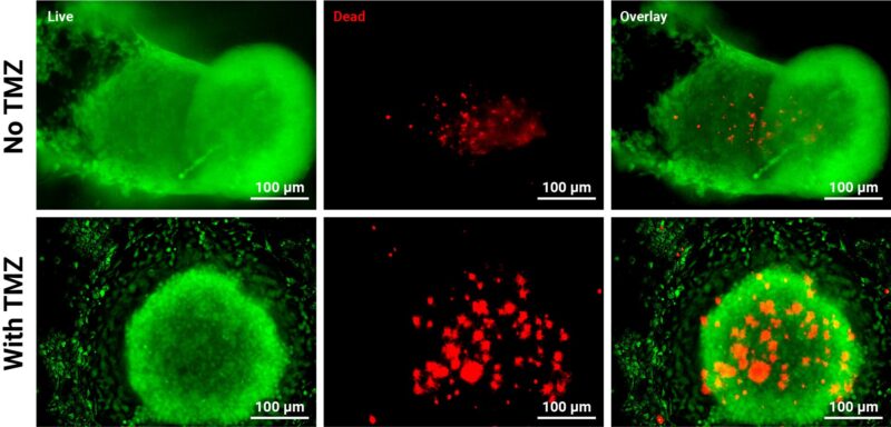
Figure 2: GBM tumoroids are susceptible to chemotherapy.
Tumoroids were grown for 3 days in VitroGel® EMT hydrogel with RocketCell™ EMT Supplement (50X), XF. The tumoroids were treated with Temozolomide (TMZ; 1mM) for 24 hours and subjected to cell viability studies.
Cyto3D® Live-Dead Assay Kit was used to label live cells (green) and dead cells (red) in the tumoroid, both with and without drug treatment. The pictures were obtained at a 20X magnification.
Resources

| GBM | Standard Kit, High-Screening Kit, Supplement only, Hydrogel only |
|---|
Related products
NEW
Culture Media and Supplements
N2-like Animal Origin-Free Supplement
New
Cell Culture Media (Animal Origin-Free)
All-in-one, animal origin-free kit for growth, expansion, and passaging of apical-out intestinal organoids.
NEW
Culture Media and Supplements
N2B27-Like Premixed Animal Origin-Free Supplement
New
Cell Culture Media (Animal Origin-Free)
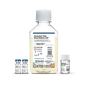
Animal origin-free complete medium for 3D and 2D neural stem cell culture
NEW
Culture Media and Supplements
B27-like, Animal Origin-Free Supplement
New

Cell Culture Media (Xeno-Free)
High-performance, xeno-free kit for superior expansion of human mesenchymal stem cells (hMSC)