Research Highlights
3D Hydrogel Embedding Boosts the Immunomodulatory Power of human gingiva-derived Stem Cells: A New Strategy for Regenerative Medicine
VitroGel® MSC hydrogel enhances anti-inflammatory response in human gingiva-derived mesenchymal stromal cells (hG-MSCs), offering a promising approach to optimize cell-based therapies for inflammatory diseases.
Category:
3D Cell Model
Subategory:
In vitro model
Cell Types:
Human gingiva-derived mesenchymal stromal cells
(hG-MSCs)
Hydrogel:
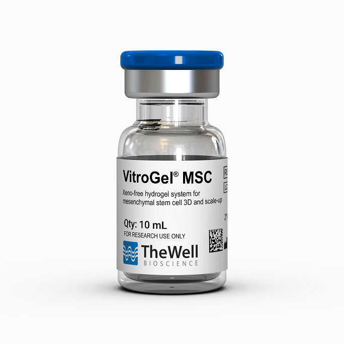
VitroGel® MSC (VHM03)
Institutions:
Competence Center for Periodontal Research, University Clinic of Dentistry, Medical University of Vienna, Vienna, Austria
Team:
Katharina Schwarz, Oliwia Miłek, Merjem Bećirović, Christian Behm, Karl Heinrich Schneider & Oleh Andrukhov
Mesenchymal stromal cells (MSCs) have emerged as promising candidates for regenerative medicine due to their remarkable ability to modulate the immune system and promote tissue repair. These cells can be isolated from various tissues, including dental sources like gingiva (gum tissue), which is particularly advantageous as it is easily accessible and often discarded as clinical waste. However, despite promising results in laboratory studies, clinical applications of MSC-based therapies have faced significant challenges, particularly low cell survival and restricted retention following patient transplantation. This critical bottleneck has hindered the translation of MSC therapies from bench to bedside. The conventional method of growing cells in 2D culture dishes fails to mimic the complex 3D environment cells naturally experience in the body, potentially compromising their therapeutic functions. Without a supportive 3D microenvironment, transplanted MSCs often fail to maintain their immunomodulatory capabilities, limiting their effectiveness in treating inflammatory conditions such as periodontitis, bone defects, and other tissue disorders.
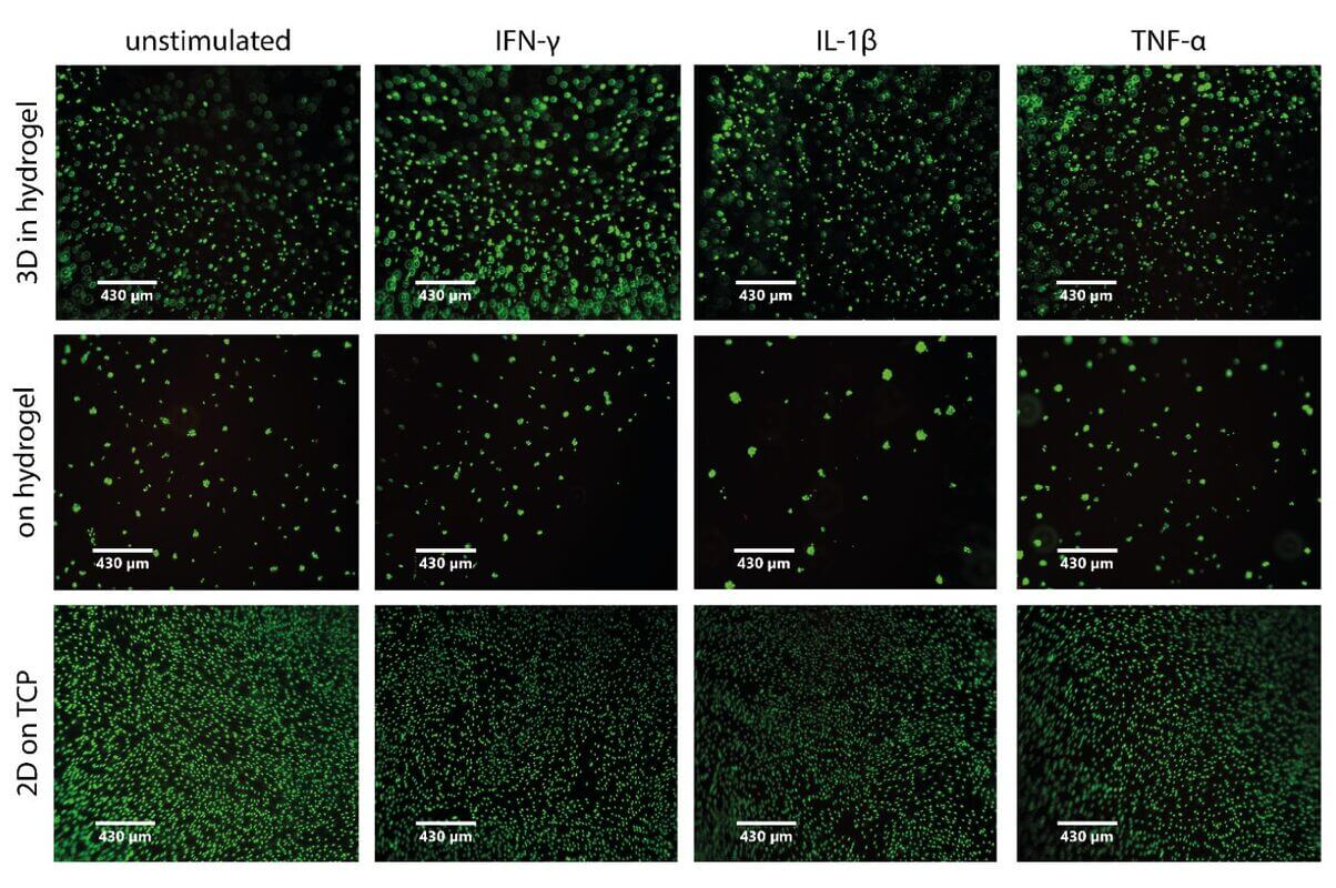
To address these challenges, researchers investigated whether embedding human gingiva MSCs (hG-MSCs) in the VitroGel® MSC hydrogel could enhance their immunomodulatory properties compared to conventional 2D cultures. The study compared three culture conditions: cells embedded within the hydrogel (3D), cells cultured on top of the hydrogel, and cells grown on standard tissue culture plastic (2D). Under both resting conditions and in the presence of pro-inflammatory cytokines (IFN-γ, IL-1β, and TNF-α), the researchers measured metabolic activity, cell viability, and the expression of key immune mediators, including IL-8, COX-2, PGE2, IDO-1, PD-L1, PD-L2, and TSG-6 (Figure 1). VitroGel® MSC hydrogel enabled the creation of a physiologically relevant 3D microenvironment that significantly altered cellular behavior. Notably, while metabolic activity was reduced in 3D-embedded cells likely due to diffusion limitations within the hydrogel, cell viability remained largely preserved. More importantly, the 3D environment dramatically enhanced the expression of several anti-inflammatory mediators: COX-2 and PGE2 were consistently upregulated, TSG-6 expression was significantly elevated across all conditions, and the hydrogel embedding modulated the expression of immune checkpoint molecules, PD-L1 and PD-L2, in a cytokine-dependent manner.
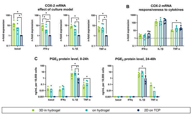
Figure 1: COX-2 and PGE2 expression in response to 3D-embedded hG-MSCs exposed to resting and pro-inflammatory cytokine conditions, demonstrating that hydrogel encapsulation enhances the anti-inflammatory and immunomodulatory potential of hG-MSCs.
This study provides compelling evidence that the 3D microenvironment created by VitroGel® MSC fundamentally reshapes the immunomodulatory profile of gingiva-derived MSCs, enhancing their anti-inflammatory and tissue-protective functions. By embedding hG-MSCs in this soft hydrogel, researchers were able to promote a more therapeutically favorable phenotype, characterized by elevated expression of key mediators like PGE2 and TSG-6 that are crucial for resolving inflammation and promoting tissue regeneration. These findings underscore the critical importance of 3D culture models in optimizing MSC-based therapies and suggest that VitroGel® MSC could serve as an effective delivery system to improve cell survival, retention, and function after transplantation.
The study also highlights the potential of hydrogel-based approaches to tailor stem cell responses to specific inflammatory environments, offering a versatile platform for personalized regenerative medicine strategies. Future research building on these findings, including co-culture experiments with immune cells and in vivo validation studies could pave the way for more effective treatments for inflammatory diseases, periodontal disorders, and craniofacial regeneration, ultimately bridging the gap between promising laboratory results and successful clinical outcomes.
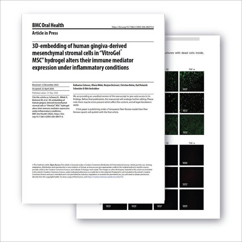
Read the publication on Springer Nature
RELATED PRODUCTS
Discover how VitroGel® can support your cell culture research
Explore our updated resources for a concise overview of VitroGel®’s key features and application areas.
Comparison of VitroGel® vs. Animal-based ECM
Discover the 20+ advantages of VitroGel® over animal-based ECM with this comprehensive comparison on key features, operation, application, and storage conditions.
Learn More
What is VitroGel® | Overview Video
Learn why VitroGel® is the leading animal-free hydrogel for 3D cell culture.
Learn More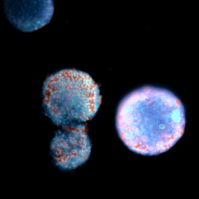
End-to-end Platform for Stem Cell Culture
Learn about the xeno-free platform to simplify and streamline stem cell research.
Learn MoreComplete Guide for Xeno-Free Stem Cell Culture: Bridge 2D and 3D culture from end-to-end.
This comprehensive product guide features validated applications, optimized workflows, and integrated solutions designed to support reproducible cell expansion, maintenance, and differentiation across a wide range of stem cell research applications.