Uncategorized
Culture Apical-Out Organoids with Xeno-Free VitroGel® System – Long Version
Introduction
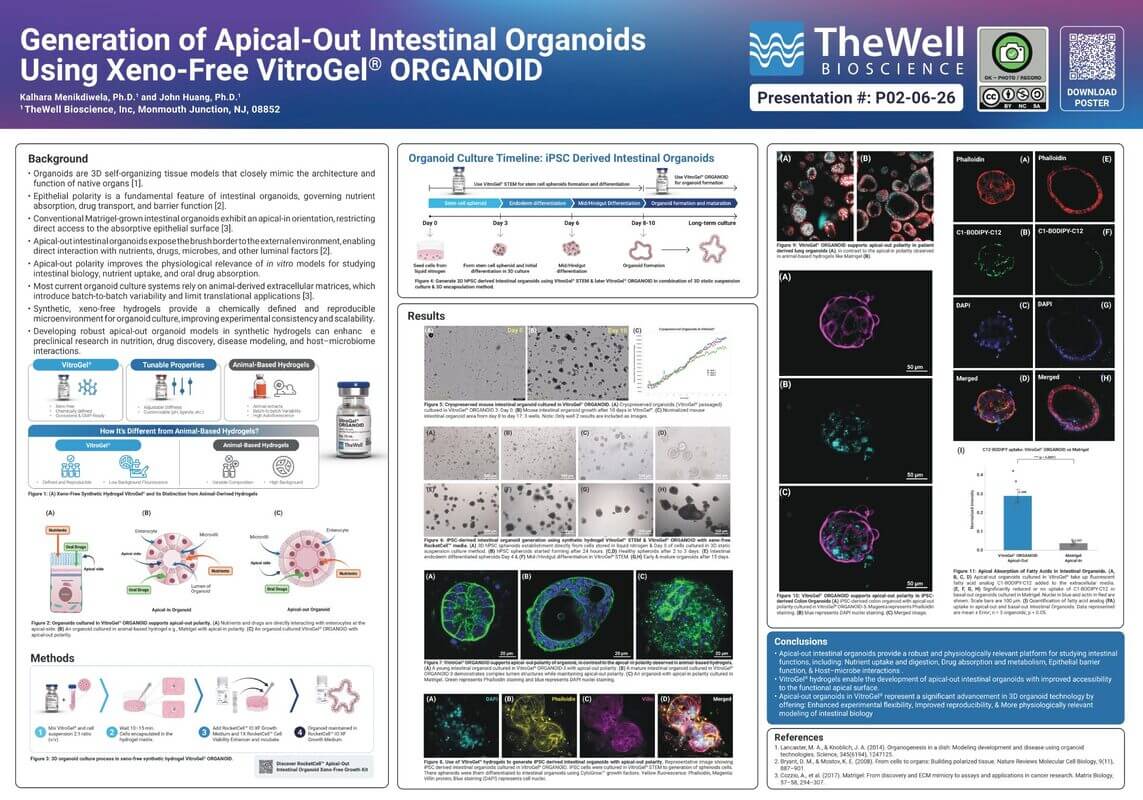
Organoids are three-dimensional (3D) miniature, self-organized tissue structures that recapitulate the functional and structural features of organs in vivo. These in vitro models exhibit greater physiological similarity to intact tissues compared to traditional two-dimensional (2D) cell cultures, making them invaluable tools for studying developmental biology, disease mechanisms, drug screening, and personalized medicine [1]. While numerous factors regulate organoid functionality, the polarity of organoids plays a crucial role in their development and functions. Polarity, which is the asymmetric distribution of cellular components, mirrors tissue organization in vivo, allowing for the study of complex biological processes like epithelial barrier function, nutrient uptake, and host-microbiome interactions [2].
Most organoid cultures currently depend on animal-derived extracellular matrices (ECMs), like Matrigel, to support growth and differentiation [3]. While these systems have advanced research, they come with key limitations. Organoids grown in such matrices often develop an apical-in polarity, where the apical side of epithelial cells faces the interior (apical-in polarity), and the basal side interacts with the surrounding matrix (Figure 1), limiting the acess to the apical side to study epithilial-nutrient interaction and uptake, drug interactions, or host-microbiome studies [1]. Unfortunately, the direction of interaction and absorption of such organoids is opposite to what we see naturally, as it occurs at the basal side. Thus, apical-in organoids (cultured in animal-based hydrogels) fail to mimic and recapitulate the interactions we see in vivo. While the apical side of the organoid can be accessible via expensive and labor-intensive approaches (e.g., microinjection), such techniques have notable drawbacks. Recent attempts to generate organoids with apical-out polarity, where the apical surface faces outward, have involved transferring organoids from animal-based ECM into suspension culture or using diluted hydrogel systems [4, 5]. Moving organoids out of ECM like Matrigel and into suspension culture can disrupt cell-matrix interactions, leading to instability, loss of function, and reduced viability [4, 5].

Figure 1. Organoids cultured in VitroGel® Organoid supports apical-out polarity. (A) Nutrients and drugs are directly interacting with enterocytes at the apical side. (B) An organoid cultured in animal-based hydrogel, e.g., Matrigel, with apical-in polarity. (C) An organoid cultured in VitroGel® ORGANOID with apical-out polarity.
In addition to polarity issues, animal-derived ECMs have disadvantages like batch-to-batch variability, potential immunogenicity, and xenogeneic components, which limit their clinical applications [3]. Synthetic, xeno-free hydrogels have been proposed as alternatives, but few can fully support organoid growth and differentiation across various culture methods and organ types [5, 6]. Long-term culture of organoids—lasting months—remains challenging in animal-based ECM systems due to uncontrolled growth patterns that may mimic tumorigenesis rather than stable tissue maintenance [3].
VitroGel® ORGANOID is a synthetic, xeno-free hydrogel system designed to support the full growth and differentiation of organoid cultures. Unlike animal-derived matrices such as Matrigel, VitroGel® eliminates batch-to-batch variability by providing a reproducible, chemically defined environment. In addition, VitroGel® can be handled at room temperature, making it well-suited for organoid scale-up and laboratory automation. A key advantage of VitroGel® is its ability to support the growth and passages of organoid with the natural apical-out polarity. Single cells or tissue fragments can be directly embedded in VitroGel® to generate mature organoids with apical-out orientation. Beyond polarity, VitroGel® hydrogel also enables long-term organoid survival — sustaining cultures for over 60 days while preserving both morphological and structural integrity. When combined with the RocketCell™ Apical-Out Intestinal Organoid Xeno-Free Growth Kit, TheWell Bioscience provides a seamless, fully defined workflow of the VitroGel® hydrogel system for apical-out intestinal organoid expansion.
In this white paper, we discuss: (1) Methods for expanding and culturing organoids with apical-out polarity using VitroGel® hydrogels. (2) Strategies for organoid passaging and long-term maintenance. (3) The ability of VitroGel® to support seamless transitions between different hydrogel systems without compromising organoid structure or function.
Materials
- VitroGel® ORGANOID-3 (Cat. No: VHM04-3)
- RocketCell™ Organoid Xeno-Free Essential-Core Medium (Cat. No.: RC04-OCM)
- CytoGrow™ Growth Factors
- EGF (Human), Recombinant Protein, CHO, Tag Free (Cat. No: CG014)
- Noggin (Human), Recombinant Protein, CHO, Tag Free (Cat. No: CG059
- R-Spondin1 (Human), Recombinant Protein, CHO, Tag Free (Cat. No: CG067)
- RocketCell™ Apical-Out Intestinal Organoid Xeno-Free Growth Kit (Cat. No.: RCO3-GK)
- VitroGel® ORGANOID-5
- RocketCell™ IO XF Cell Suspension Medium
- RocketCell™ IO XF Growth Medium
- RocketCell™ Cell Viability Enhancer (1000X)
5. Intestinal organoid fragments
6. Culture Vessels
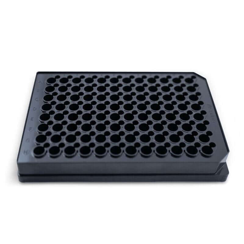
- VitroPrime™ Spread-Attach plates for the encapsulation method, 96-well (Cat. No.: VP-SA96W)
- VitroPrime™ 3D Culture and Imaging Plate, 96-well (Cat. No.: VP-3D96W5)
7. Animal-based hydrogel (Matrigel)
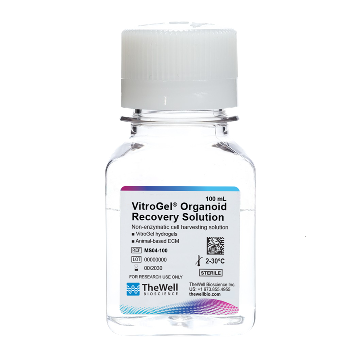
8. VitroGel® Organoid Recovery Solution (Cat. No.: MS04)
9. DPBS (Wash buffer, no calcium, no magnesium)
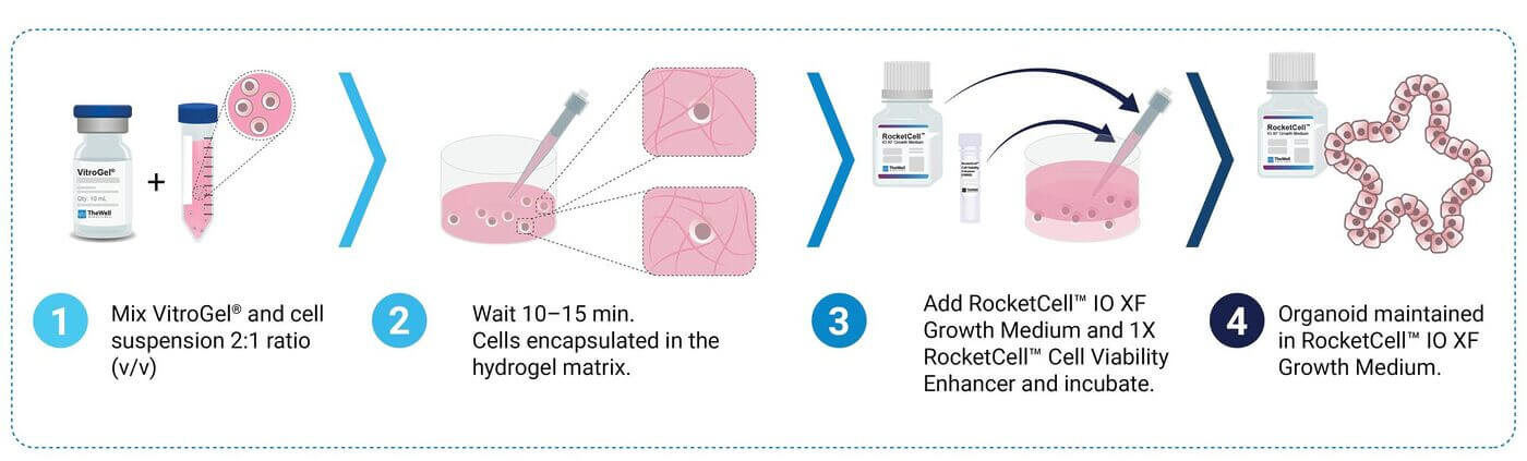
Figure 2. 3D Organoid Culture Process
Methods
3D Organoid Culture Process*: Short-term & Long-term Culturing in VitroGel®
Intestinal organoids were used in this experiment. Cryopreserved organoids were cultured in Matrigel and VitroGel® hydrogel by using RocketCell™ Apical-out Intestinal Organoid Xeno-Free Growth Kit (Cat. No. RC03-GK) (Figure 2).
1. For VitroGel®, a suspension of organoid fragments or cells was prepared with RocketCell™ IO XF CS Medium (cell suspension medium).
Please Note: Recommended to add 3X RocketCell™ Cell Viability Enhancer before mixing with gel. For example, when resuspending a cell pellet in 1000 mL of Suspension Medium, add 3 μL of RocketCell™ Cell Viability Enhancer. Scale as needed.
2. VitroGel® ORGANOID and organoid suspension were mixed at 2:1 v/v ratio and cover the well bottom (Encapsulation method: Figure 2) and kept for 15 minutes at room temperature for a soft gel formation. Similarly, organoids suspended in Matrigel were incubated for 15 minutes as domes in an incubator at 37°C for gel formation.
3. Once ready, organoids in both VitroGel® & Matrigel were treated with RocketCell™ IO XF G Medium (growth medium) with 1X RocketCell™ Cell Viability Enhancer for the first 24 to 48 hours. The media is supplemented with essential growth factors / inhibitors for 24 to 48 hours to stimulate organoid growth, and a cell viability enhancer will provide initial protection.
4. Organoid cultures were then maintained in RocketCell™ IO XF G medium (growth medium) without RocketCell™ Cell Viability Enhancer.
5. Short-term culturing, changing media every 2-3 days. Organoids start maturing after 8-10 days on RocketCell™ IO XF G Medium (growth medium). Mature organoids maintained in RocketCell™ IO XF G Medium for 10 to 15 days can be used for expansion/passaging or any other downstream application.
6. Long-term culturing, similar to short-term culturing, media was changed every 2-3 days. Organoids can be maintained in the VitroGel® hydrogel system on RocketCell™ IO XF G Medium for over 60 days.
*Please refer to  VitroGel® ORGANOID Protocol for more information and detailed protocol.
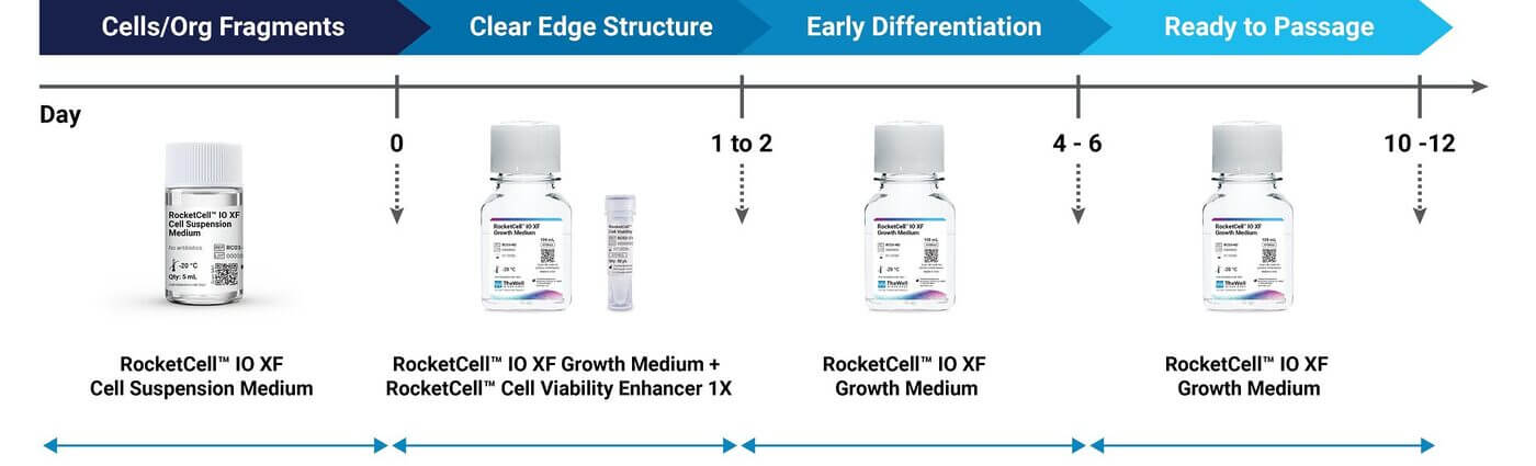
Figure 3. Intestinal organoid expansion in VitroGel® hydrogel system.
Organoid expansion/passaging steps in VitroGel® (Figure 3)
1. Harvest organoids cultured in VitroGel® hydrogel system or in animal-based gel using VitroGel® Organoid Recovery solution.
- Warm the VitroGel® Organoid Recovery Solution to 37°C.
- Take organoids out of the incubator and remove the medium covering the top of the hydrogel. Wash the hydrogel two times with DPBS.
- Add 1 mL warm VitroGel® Organoid Recovery Solution to the well and use 1 mL a pipette to gently break the hydrogel into small pieces by gently pipetting up and down. This step can accelerate the hydrogel dissolving process.
- Add 5 mL warm VitroGel® Organoid Recovery Solution to a 15 mL conical tube and transfer the hydrogel to the tube.
Optional: Rinse the well with 1 mL warm VitroGel® Organoid Recovery Solution and combine the solution to the centrifuge tube. - Pipette the mixture up and down 2-5 times and put the tube in water/dry bath for 2-3 min. Repeat this cycle 2-3 times as required. (Optimize the pipetting times and repeat according to the gel strength and cell type).
2. Centrifuge at 100 to 350 x g for 3-5 minutes at room temperature to collect the organoid pellet. (Optimize the speed and time of centrifuge according to different cell types).
3. Make an organoid fragment or cell suspension in RocketCell™ IO XF Medium 1.
a. VitroGel® & organoid suspension were mixed at 2:1 v/v ratio. Incubate 5 minutes at room temperature for soft gel formation.
4. Cover gel with RocketCell™ IO XF Medium 2 (24 to 48hrs). Healthy organoids form clear edge structures during this time frame.
5. On day 3, replace RocketCell™ IO XF Medium 2 with RocketCell™ IO Medium 3.
6. Change media every 2-3 days & maintained on RocketCell™ IO XF Medium 3.
7. Organoids are ready to passage on day 10-12 (depending on how fast organoids grow, passaging can be done after day 5 when early differentiation is completed).
8. Harvest organoids with VitroGel® Organoid Recovery Solution (MS04) and follow step 1 through 5.
Additionally, when organoids are expanded or passaged in VitroGel® hydrogel system, they can be successfully passaged in VitroGel® systems several times while maintaining structural and morphological features unlike in most animal-based hydrogel systems. More importantly, organoids cultured in VitroGel® system can be easily passaged into other hydrogel systems including Matrigel.
*Please refer to Harvesting/Recovery Protocol for more information and detailed protocol.
A brief protocol for organoid staining
For a high-resolution organoid image, we recommend culturing organoids in VitroPrime™ 3D Cell Culture and Imaging Plate. This cover glass bottom plate supports organoid culture, fixing and staining without losing or disturbing samples for high-resolution imaging.
1. Remove the cover medium through the medium changing channel of VitroPrime™ 3D Culture and Imaging Plate.
2. Fix organoids with 4% formaldehyde solution.
3. After permeabilization & blocking treatments, organoids embedded in VitroGel® & Matrigel were incubated with primary antibodies overnight at 4°C (followed company recommended dilutions).
4. After primary antibodies are removed, wash the gel carefully 3 times with 100 μL DPBS.
5. Add an appropriate amount of secondary antibody (diluted to 1X in blocking solution) and incubate overnight at 4°C.
6. Perform immunofluorescent imaging using a confocal microscope.
*Please refer to Immunofluorescence Staining Protocol for more information and detailed protocol.
Results and Discussion
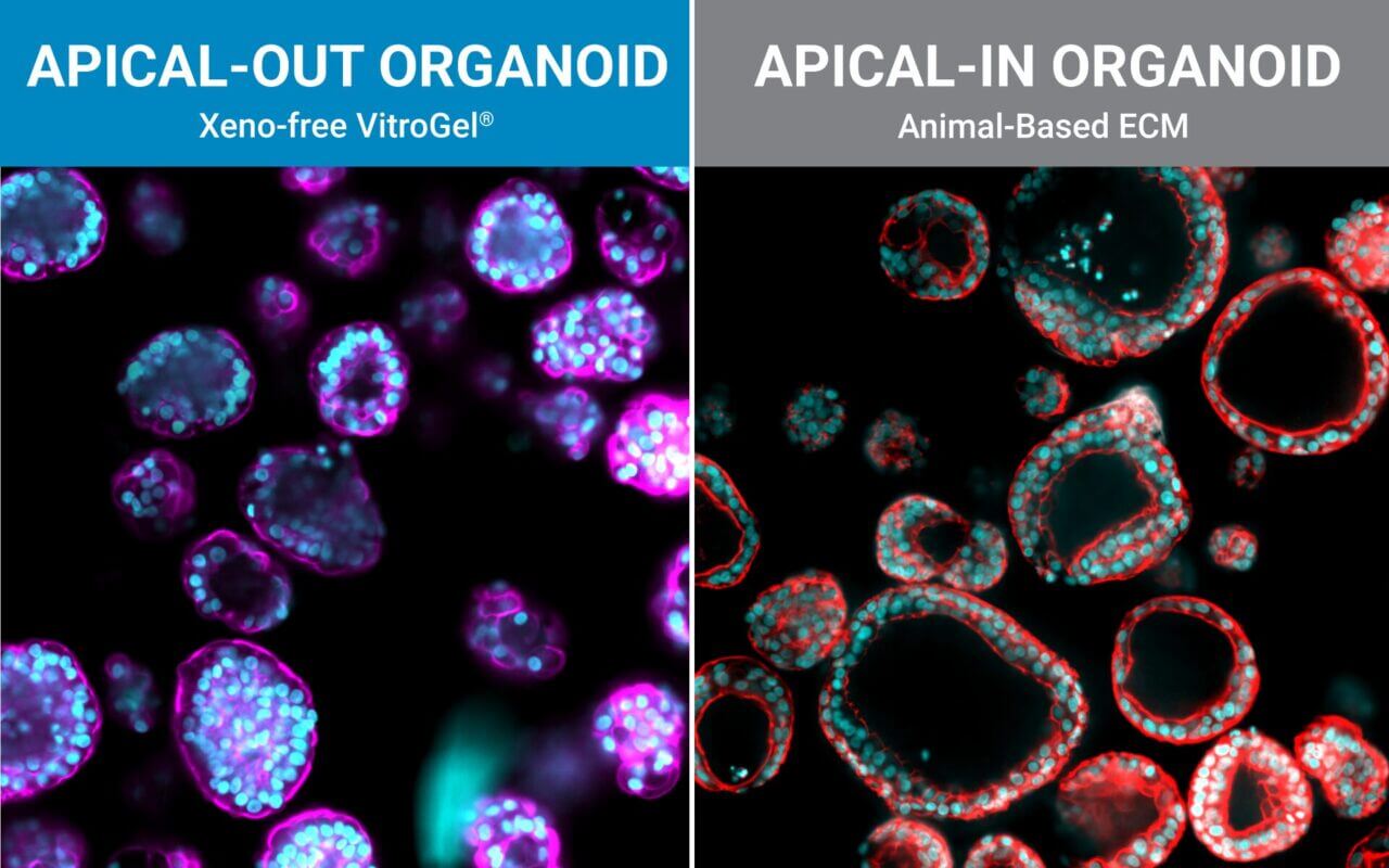
The polarity of cells and organoids plays a critical role in their development and function. We confirm the polarity of organoids cultured in both animal-based hydrogels like Matrigel as well as VitroGel® hydrogel system by performing immune staining (Figure 4A-C and Figure 5). When organoids with epithelial structures are cultured in Matrigel, the apical side of the epithelial cells typically faces inward toward the lumen, exhibiting apical-in and basal-out polarity (Figure 4C, 7D, and 7E). While this configuration supports tissue organization, it restricts experimental access to the apical surface, which is essential for studies of nutrient uptake, drug interactions, and host–microbiome dynamics [1, 4, 5, 7].
To overcome this, researchers have to either use a sophisticated microinjection approach or techniques to forcefully flip organoid polarity. However, such methods can be technically challenging and may introduce artifacts with significant drawbacks. Recent efforts to generate organoids with apical-out polarity, in which the apical surface faces outward, have typically relied on transferring organoids from animal-derived ECMs into suspension culture or by using diluted hydrogel systems [4]. However, removing organoids from supportive ECMs such as Matrigel disrupts critical cell– matrix interactions, often resulting in structural instability, loss of function, and reduced long-term viability [4, 5]. Additionally, diluted hydrogels often lack the necessary mechanical strength and bio-functional ligands, making them unsuitable for long-term culture [5]. As a result, organoids grown under these conditions may lose their physiological relevance, which is crucial for applications like disease modeling or regenerative medicine [1, 3, 5, 6].
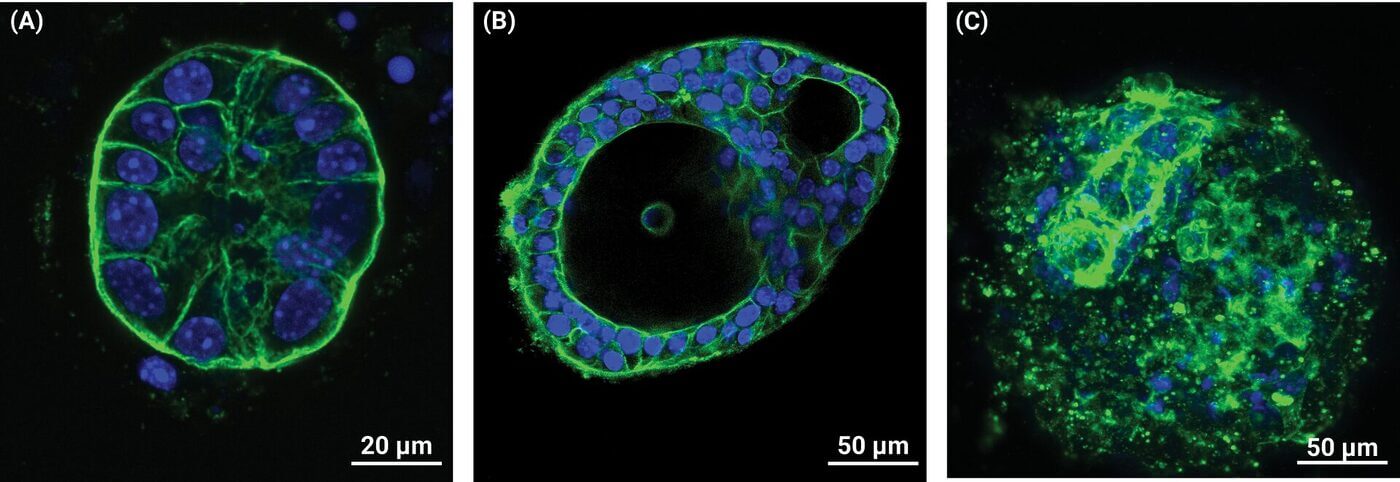
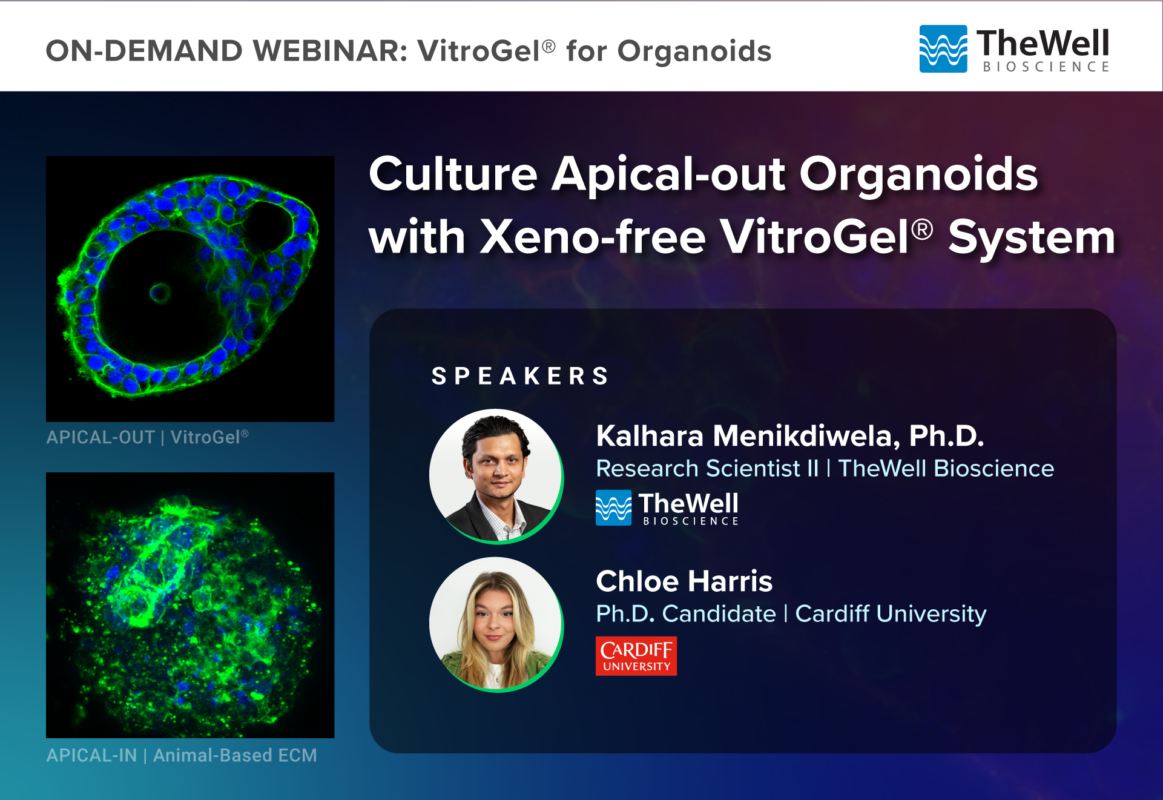
Figure 4. VitroGel® ORGANOID supports apical-out polarity of organoid, in contrast to the apical-in polarity observed in animal-based hydrogels. (A) A young intestinal organoid cultured in VitroGel® ORGANOID-3 with apical-out polarity. (B) A mature intestinal organoid cultured in VitroGel® ORGANOID-3 demonstrates complex lumen structures while maintaining apical-out polarity. (C) An organoid with apical-in polarity cultured in Matrigel. Green represents Phalloidin staining and blue represents DAPI nuclei staining.
However, when intestinal organoids are cultured in VitroGel® ORGANOID, the system naturally supports apical-out polarity, allowing direct access of the surrounding medium to the epithelial surface (Figures 4 and 5). As shown in Figure 4 and Figure 5, intestinal organoids in VitroGel® display a distinct epithelial layer facing outward (Figure 4A, 4B, and 5B), in contrast to organoids cultured in Matrigel (Figure 4C, 7D, and 7E). Remarkably, VitroGel® supports apical-out polarity even in young organoids as early as day 2 (Figure 4A).
As the organoids mature in VitroGel®, they develop lumen structures that closely recapitulate small intestinal architecture while still maintaining apical-out polarity (Figure 4B). In contrast, attempts to flip organoids from Matrigel into suspension or 2D culture often led to the collapse of the internal structure, preventing stable lumen formation. Furthermore, intestinal organoids in VitroGel® can be maintained for extended culture periods while preserving apical-out organization — a feature not achievable in traditional animal-derived hydrogel systems.
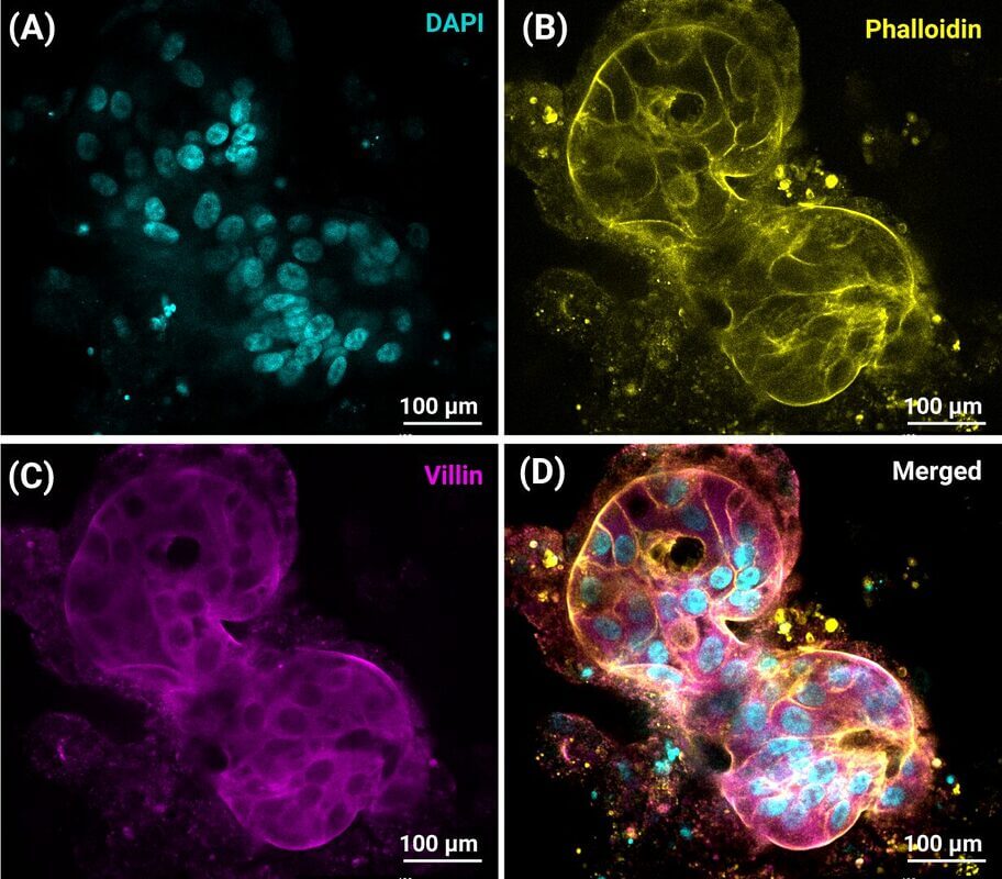
Figure 5: Use of VitroGel® hydrogels to generate iPSC-derived intestinal organoid with apical-out polarity. Representative images showing intestinal organoids cultured in VitroGel® ORGANOID. iPSC cells were culture in VitroGel® STEM to generate spheroids. Spheroids were then differentiated to intestinal organoids using CytoGrow™ Growth Factors. (A) Blue staining (DAPI) represents cell nuclei. (B) Yellow fluorescence: Phalloidin, (C) Magenta: Villin protein, (D) Merged image of the iPSC-derived intestinal organoid.
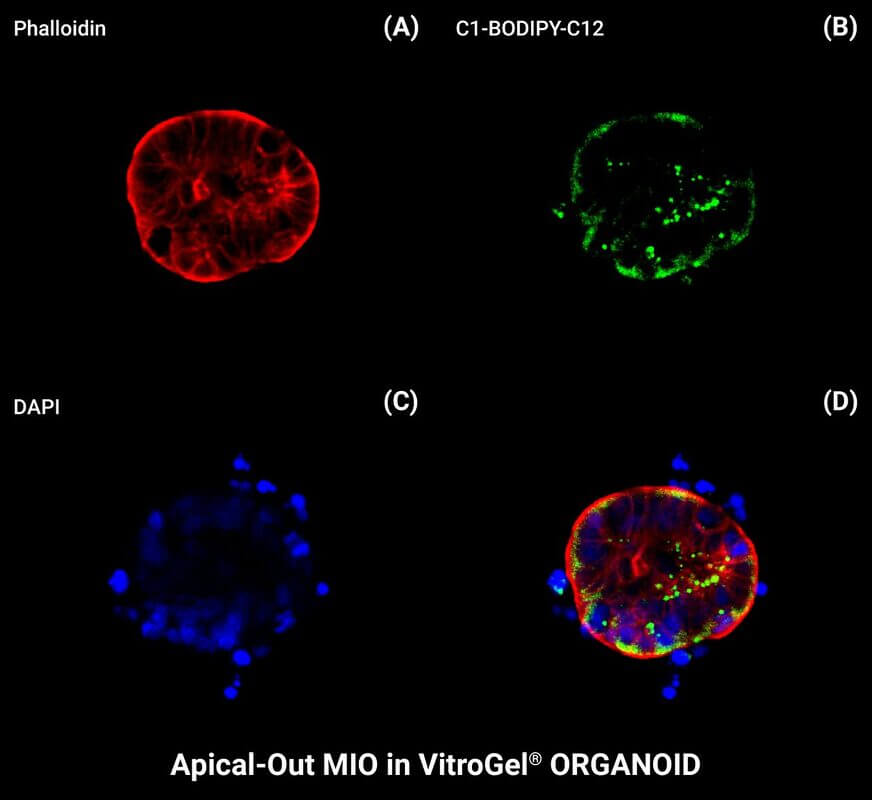
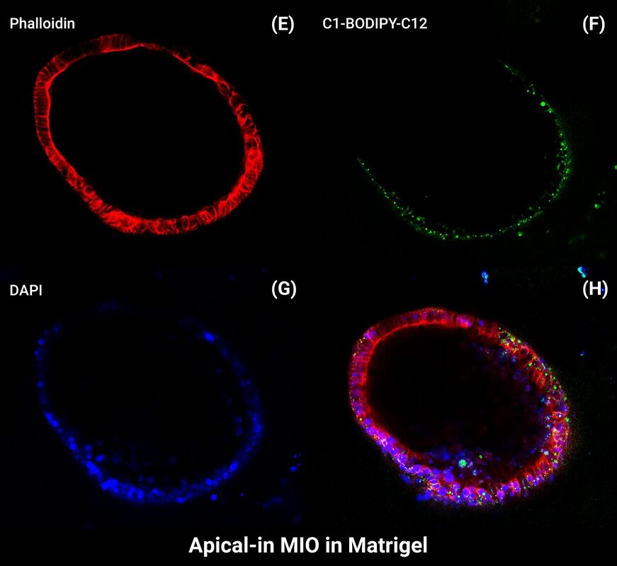
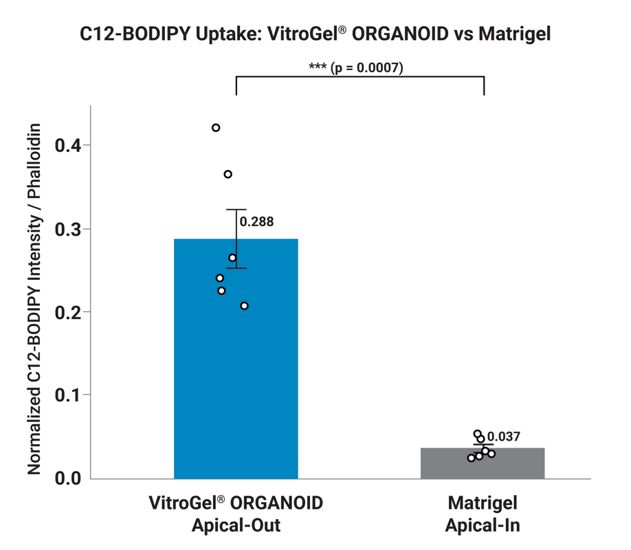
Figure 6: Apical Absorption of Fatty Acids in Intestinal Organoids. (A, B, C, D) Apical-out organoids cultured in VitroGel® ORGANOID take up fluorescent fatty acid analog C1-BODIPY-C12 added to extracellular media. (E, F, G, H) Significantly reduced or no uptake of C1-BODIPY-C12 in basal-out organoids cultured in Matrigel. Nuclei in blue and actin in red are shown. Scale bars are 100 µm. (I) Quantification of fatty acid analog (FA) uptake in apical-out and basal-out intestinal organoids. Data represented are mean + Error; n=6 organoids ; p < 0.05.
To further confirm the polarity of intestinal organoids cultured in VitroGel® and Matrigel, the cultures were treated with the fluorescent fatty acid analog C1-BODIPY-C12 (BODIPY-labeled dodecanoic/lauric acid), a functional probe for epithelial polarity. In the native intestine, long-chain fatty acids are absorbed through the apical (brush border) membrane of enterocytes. Because BODIPY-C12 mimics this process, it is efficiently taken up only when the apical surface is exposed to the culture medium. Thus, uptake of BODIPY-C12 provides a functional readout of organoid orientation, distinguishing apical-out organoids, where the apical membrane faces the external medium (as observed in VitroGel®), from apical-in organoids, where the apical surface is enclosed within the lumen (as typically seen in Matrigel).
As indicated in Figure 6 a clear BODIPY-C12 uptake (green staining) into the organoid lumen was observed in intestinal organoid cultured in VitroGel® ORGANOID compared to Matrigel-cultured organoids (Figure 6B vs. 6F). In contrast to VitroGel®-cultured organoids, no detectable BODIPY-C12 uptake was observed in organoids cultured in Matrigel. When normalized to red (structural/membrane marker Phalloidin) and compared at the wall, VitroGel® apical-out organoids showed a mean ratio of 0.298 ± 0.064 (SEM), roughly 8-fold higher than Matrigel apical-in organoids at 0.036 ± 0.009. This is consistent with organoid polarity directly controlling lipid accessibility.
In apical-out organoids, the absorptive apical membrane faces outward into the culture medium, giving BODIPY-C12 direct access — mimicking physiological dietary fat absorption at the intestinal brush border.
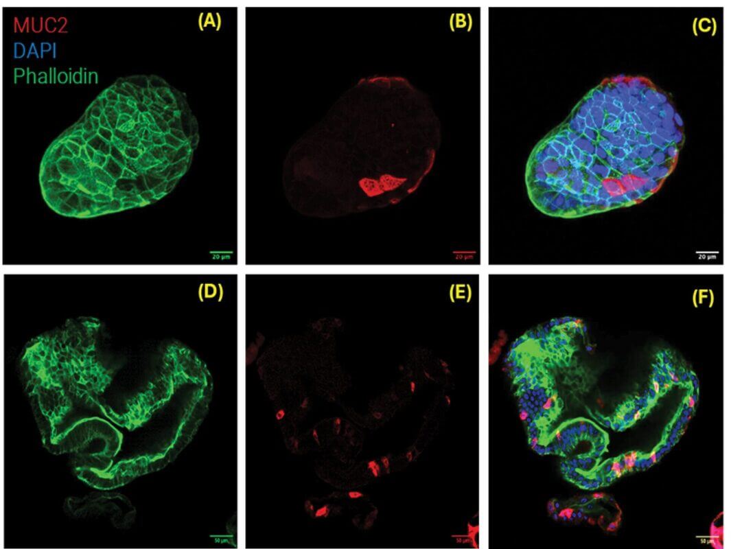
Figure 7: VitroGel® ORGANOID supports apical-out polarity colorectal cancer (CRC) organoids (A-C), in contrast to the apical-in polarity observed in animal-based hydrogels (D-F). (A) CRC organoid cultured in VitroGel® ORGANOID-3 with apical-out polarity. Green indicates epithelial staining with Phalloidin staining. (B) A mature CRC organoid cultured in VitroGel® ORGANOID-3, and MUC2 staining represents goblet cells. (C) Merged image of CRC organoid cultured in VitroGel® ORGANOID with apical-out polarity. (D) A mature CRC organoid with apical-in polarity cultured in Matrigel. Green indicates epithelial staining with Phalloidin staining. (E) MUC2 staining represents goblet cells. (F) Merged image of CRC organoid cultured in Matrigel with apical-in polarity. [Research and image credit: Chloe Harris at Cardiff University carried out this work and staining. Images were reprocessed by Kalhara Menikdiwela at TheWell Bioscience.]
In apical-in (Matrigel) organoids, the apical surface faces the enclosed lumen, so BODIPY-C12 in the medium first has to cross the basolateral membrane or diffuse in through a restricted opening, substantially limiting uptake. The much lower and more consistent Matrigel values (tight SEM) versus the higher, more variable VitroGel® values also fit this: apical-out access is a more “open” absorption route, while apical-in access is more indirect and diffusion-limited.
Furthermore, colorectal cancer (CRC) organoids and patient-derived lung organoids can also be cultured in VitroGel®, where they naturally develop an apical-out polarity (Figures 7 and 8). This unique structural orientation allows the apical surface of CRC and lung organoids to be directly exposed to the surrounding culture environment while maintaining a physiologically relevant 3D culture system.
Figure 7 demonstrates mature CRC organoids cultured in VitroGel® with apical-out polarity. As the CRC organoids mature in VitroGel®, they retain their apical-out orientation while developing structural and morphological features that closely resemble native tissue architecture (Figure 7A–C). Similarly, airway lung organoids cultured in VitroGel® exhibited apical-out polarity, in contrast to the conventional apical-in polarity typically observed in animal-derived matrices (Figure 8).
This apical-out configuration significantly simplifies experimental workflows by reducing or eliminating the need for technically challenging procedures, such as microinjection, that are often required for traditional apical-in organoid models. As a result, VitroGel®-supported apical-out CRC and lung organoid models provide a powerful, scalable, and physiologically relevant platform for translational research, personalized medicine, drug discovery, host–pathogen interaction studies, and advanced in vitro disease modeling.
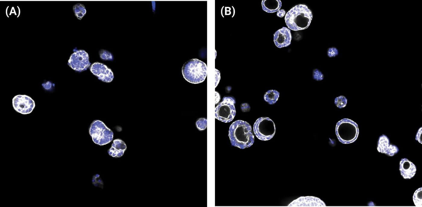
Figure 8: VitroGel® ORGANOID supports apical-out polarity in patient-derived lung organoids (A), in contrast to the apical-in polarity observed in animal-based hydrogels like Matrigel (B).
VitroGel® ORGANOID supports organoid formation from a wide range of sources, including patient-derived samples, established cell lines, and iPSC-derived organoid fragments. The system enables the natural generation of apical-out organoids starting from either single cells or small cell clusters, providing a robust and versatile platform for diverse organoid applications. As shown in Figure 9, stem cell–containing clusters cultured in the VitroGel® ORGANOID system developed into mature intestinal organoids within 12 days. A well-defined outer structure was evident as early as 24 to 72 hours (3 days), with progressive organoid formation observed by day 6 (Figure 9A, B). When cultures were maintained in RocketCell™ IO XF G Medium for over 12 days, fully developed mature organoids were obtained (Figure 9C).

Figure 9. VitroGel® ORGANOID supports intestinal organoid generation out of cell clusters. (A) Cell clusters develop clear-edged structures within 72 hours (3 days) in VitroGel® ORGANOID. (B) Healthy young organoids form in 6 days. (C) Mature intestinal organoids in VitroGel® ORGANOID.
Unlike animal-derived ECM systems, VitroGel® ORGANOID maintains organoid integrity and functionality over extended periods, making it highly suitable for long-term culture. One of VitroGel’s key advantages is its ability to sustain organoid growth for months, whereas animal-based ECMs often fail due to uncontrolled growth patterns. The undefined and variable composition of hydrogels such as Matrigel or BME, combined with the apical-in polarity they promote, frequently results in the accumulation of dead cells and toxic byproducts within mature organoids, limiting culture longevity.
To evaluate the capacity of VitroGel® ORGANOID to support long-term organoid growth, we maintained intestinal organoid cultures for more than two months and compared them with parallel cultures in Matrigel (Figure 10). Organoids grown in VitroGel® ORGANOID-3 demonstrated stable and controlled growth for over 60 days (Figure 10). In contrast, organoids cultured in Matrigel exhibited rapid and uncontrolled growth, resembling a tumor-like pattern, with most organoids rupturing and dying after 30–40 days, likely due to toxicity buildup within the mature structures (Figure 10B).
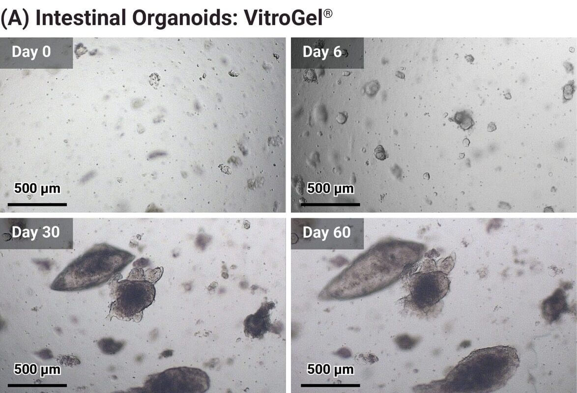
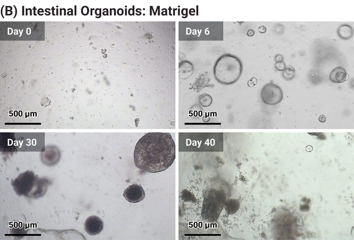
Figure 10: VitroGel® Supports Long-term Organoid Growth. (A) Bright-field images of intestinal organoids cultured and maintained in VitroGel® ORGANOID-3 over 60 days in RocketCell™ Apical-Out Intestinal Organoid Xeno-Free Growth Kit (from day 0 through day 60). (B) Intestinal organoids cultured and maintained in Matrigel with RocketCell™ Apical-Out Intestinal Organoid Xeno-free Growth Medium. Organoids in Matrigel started dying after 30 days in 3D culture.
In order to further validate the ability of VitroGel® ORGANOID to support long-term organoid growth while preserving functional and structural features, intestinal organoids were stained with lineage-specific markers (Figure 11). Organoids maintained in VitroGel® ORGANOID-3 for 50 days retained their structural integrity over time. ZO-1, a tight junction protein expressed in the intestinal epithelium, plays a critical role in forming the barrier that regulates intestinal wall permeability. Clear ZO-1 expression was observed in mature organoids cultured in the VitroGel® system (Figure 11).
In addition, β-catenin—a central component of the Wnt/β-catenin signaling pathway and an essential regulator of intestinal homeostasis, stem cell renewal, and tissue regeneration— was also positively expressed (red) in these mature intestinal organoids. Robust β-catenin expression further demonstrates the healthy growth and maintenance of intestinal functionality over long-term culture in VitroGel® ORGANOID (Figure 11).
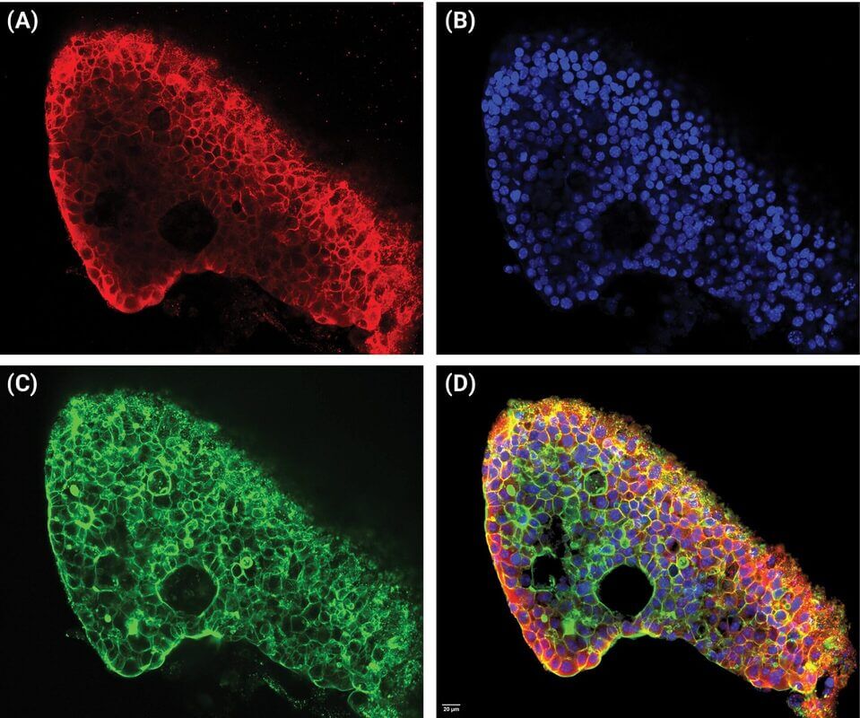
Figure 11: VitroGel® ORGANOID supports long term organoid growth while maintaining structural integrity. (A) A mature intestinal organoid cultured in VitroGel® ORGANOID stained with β-catenin (in red). (B) DAPI nuclei staining (in blue). (C) Intestinal organoid cultured in VitroGel® ORGANOIDS stained with ZO-1 (in green). (D) merged image.
The VitroGel® system not only maintains and supports apicalout polarity, but also enables stable passaging of organoids for expansion studies (Figure 11A). This capability allows researchers to establish their own apical-out organoid banks within a 100% synthetic, xeno-free VitroGel® system. As shown in Figure 11A, intestinal organoids were successfully passaged multiple times in VitroGel® while maintaining structural integrity and apical-out orientation. Organoids can be reliably passaged multiple times (over six passages) in the VitroGel® ORGANOID system, while consistently maintaining their structural and morphological integrity throughout successive generations.
Meanwhile, if researchers wish to compare both apical-out and apical-in structures from the same organoid source, organoids maintained in the VitroGel® system can be readily transferred into other hydrogel systems (such as Matrigel). Notably, organoids pre-cultured in VitroGel® demonstrate enhanced adaptability and faster stabilization in the new microenvironment compared to those generated directly in animal-based matrices. Figure 11B demonstrates that organoids transferred from VitroGel® to Matrigel recovered steadily and exhibited stable growth, further confirming the advantages of VitroGel® synthetic hydrogels over other commercially available hydrogel systems.
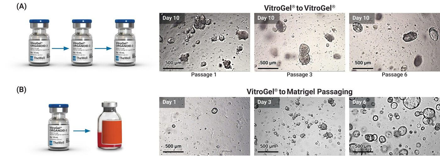
Figure 12: VitroGel® Supports Organoid Expansion & Passaging. (A) Intestinal organoids were expanded and passaged 6 times within the VitroGel® ORGANOID-3 hydrogel system. (B) Intestinal organoids grown in VitroGel® ORGANOIDS were expanded into Matrigel hydrogel system with smooth recovery and organoid growth.
Conclusion
VitroGel® synthetic hydrogel system offers a robust, reproducible, and scalable platform for organoid culture. Being a fully synthetic and chemically defined hydrogel, it ensures consistent composition and minimal batch-to-batch variability, which is critical for reproducible experiments. Its mechanical and biochemical properties can be finely tuned to mimic specific tissue microenvironments, providing better control over organoid development.
Organoids cultured in VitroGel® with RocketCell™ Apical-Out Intestinal Organoid Xeno-Free Growth Media can be easily passaged multiple times while maintaining structural integrity and viability, and they can rapidly adapt when transferred to other hydrogel systems, offering flexibility for comparative studies but also suitable for organoid banking. Unlike Matrigel, which is animal-derived and contains residual growth factors, VitroGel® is xeno-free and minimizes background signaling, making it more suitable for translational research and clinical applications. In addition, VitroGel® can be handled at room temperature, making it well-suited for organoid scale-up and laboratory automation.
More importantly, VitroGel® supports apical-out polarity, long-term culture, and diverse organoid types, representing a key advancement for advanced research and therapeutic applications. Together, these features make VitroGel® ORGANOID a superior platform for reliable, scalable, and precise organoid culture.
Read the White Paper
Fill out the form below to explore the data, imaging results, and culture methods that demonstrate how synthetic hydrogels can simplify organoid research while improving reproducibility and scalability.
Products Used
Organoid Development


CytoGrow™ Growth Factors
EGF, Noggin, R-Spondin1
Organoid Recovery
Organoid Analysis
References:
- Lancaster, M. A., & Knoblich, J. A. (2014). Organogenesis in a dish: Modeling development and disease using organoid technologies. Science, 345(6194), 1247125.
- Bryant & Mostov, (2008). From cells to organs: building polarized tissue” — Nature Reviews Molecular Cell Biology (2008)
- Cozzio, A., et al. (2017). Matrigel: From discovery and ECM mimicry to assays and applications in cancer research. Matrix Biology, 57-58, 294-307.
- Li, Y., et al. (2020). Generating apical-out intestinal organoids by suspension culture for microbiome studies. Nature Protocols, 15(3), 1234-1256.
- Gjorevski, N., et al. (2016). Designer matrices for intestinal stem cell and organoid culture. Nature, 539(7630), 560-564.
- Cruz-Acuña, R., et al. (2017). Synthetic hydrogels for human intestinal organoid generation and colonic wound repair. Nature Cell Biology, 19(11), 1326-1335.
- Sachs, N., et al. (2019). Long-term expanding human airway organoids for disease modeling. EMBO Journal, 38, e100300.